Sample Outputs and Deliverables
This page shows the concrete outputs that sponsors, CROs, and investigators receive from each acne severity assessment. The AI generates standardised reports immediately after image submission — no central reader, no delays.
Per-visit assessment report
Each image submission generates a complete severity report, accessible to the investigator within seconds:
IGA
3
ModerateAcne Lesion And Density Index
3.5
ModerateScale 0 – 4 (continuous)
Body site
Left diagonal
Image quality
92%
Lesion count
36
Density
0.55
Local score
3.42
Body site
Right diagonal
Image quality
88%
Lesion count
35
Density
0.6
Local score
3.49
What the report contains
| Output | Detail |
|---|---|
| Global IGA score | 0–4 integer aligned with the FDA co-primary endpoint |
| Global ALADIN score | 0–10 continuous composite (IGA × 2.5), for secondary/exploratory use |
| Per-perspective lesion count | Individual inflammatory lesion count (papules, pustules, nodules) per half-face |
| Per-perspective density | Spatial density ratio (0–1) measuring lesion clustering |
| Per-perspective local IGA | Local severity score for each captured perspective |
| Annotated images | Each perspective with bounding boxes around detected inflammatory lesions |
| Image quality (DIQA) | Quality score per image; images below threshold are flagged for recapture |
| Timestamp | UTC timestamp of image capture and AI processing |
The bounding box overlays allow investigators to visually verify the AI's detections against their clinical observation. This audit trail supports regulatory inspection and gives sites confidence in what the system produces.
Longitudinal severity tracking
Across visits, the platform tracks severity evolution from screening to end of study:
What longitudinal tracking delivers
- Score trajectory: IGA and ALADIN at every visit, from screening to end of study
- Treatment response flag: IGA ≥ 2-grade improvement from baseline (configurable threshold)
- Absolute change: Point change in IGA and ALADIN vs. baseline at each visit
- Visual chart: Graphical severity curve across all timepoints
This enables investigators and sponsors to identify responders and non-responders during the trial — not just at database lock.
Data export for EDC integration
All assessment outputs are structured for export to the sponsor's EDC system. Fields exported at each visit:
| Field | Description | Format |
|---|---|---|
| Patient identifier | Study-specific pseudonymised ID | String |
| Visit date | Date and timestamp of the assessment | ISO 8601 |
| Global IGA score | IGA severity (0–4) | Integer |
| Global ALADIN score | Composite severity (0–10) | Float |
| Global lesion count | Total inflammatory lesions (deduplicated across perspectives) | Integer |
| Per-perspective IGA | Local IGA score for each perspective | Integer |
| Per-perspective lesion count | Lesions detected per perspective | Integer |
| Per-perspective density | Spatial density per perspective | Float (0–1) |
| DIQA scores | Image quality score per perspective | Float |
| Severity classification | Clear / Almost clear / Mild / Moderate / Severe | String |
Data is transferred automatically via RESTful API or scheduled S3 export. Formats include structured JSON and CSV/Excel. Legit.Health provides IQ/OQ documentation and data mapping specifications.
AI visual outputs
Beyond scores, the AI produces visual artefacts that demonstrate exactly what the system detected and how it reached its conclusion. These are available in every per-visit report.
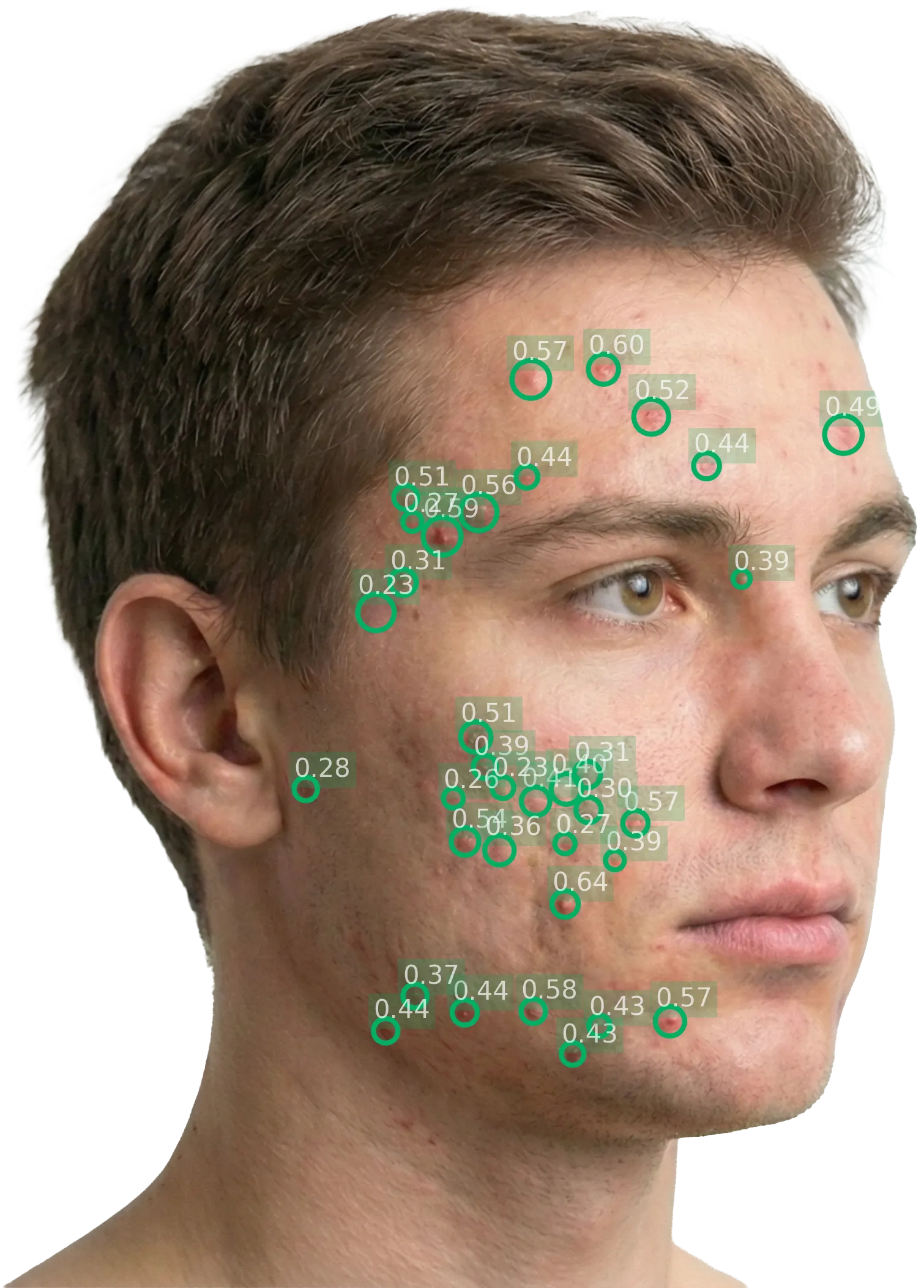
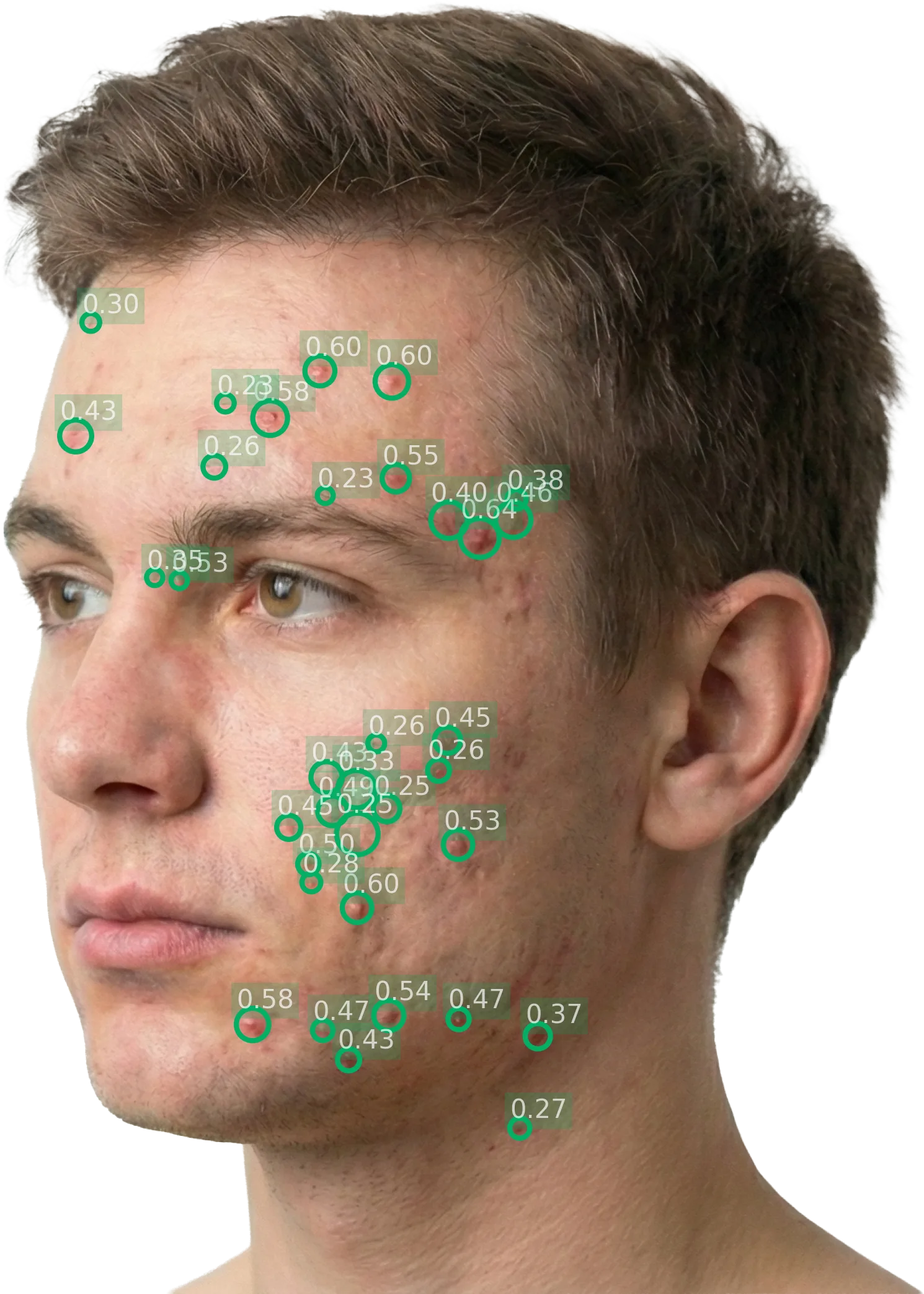
Lesion detection with bounding boxes
Each inflammatory lesion (papule, pustule, nodule) is identified and highlighted with a bounding box overlay on the original photograph. Investigators can verify the AI's detections at a glance.

Input: standardised facial photograph
Output: each detected inflammatory lesion highlighted
Facial landmark detection and pose estimation
The AI maps facial landmarks (eyes, nose, mouth, jawline) to identify the capture perspective, define region boundaries, and deduplicate lesions across overlapping perspectives.
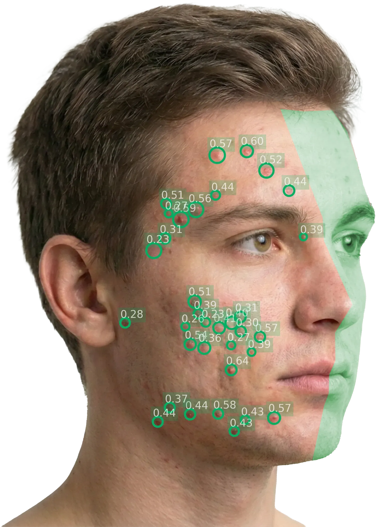
Left diagonal: the green overlay marks the region excluded from this perspective's lesion count
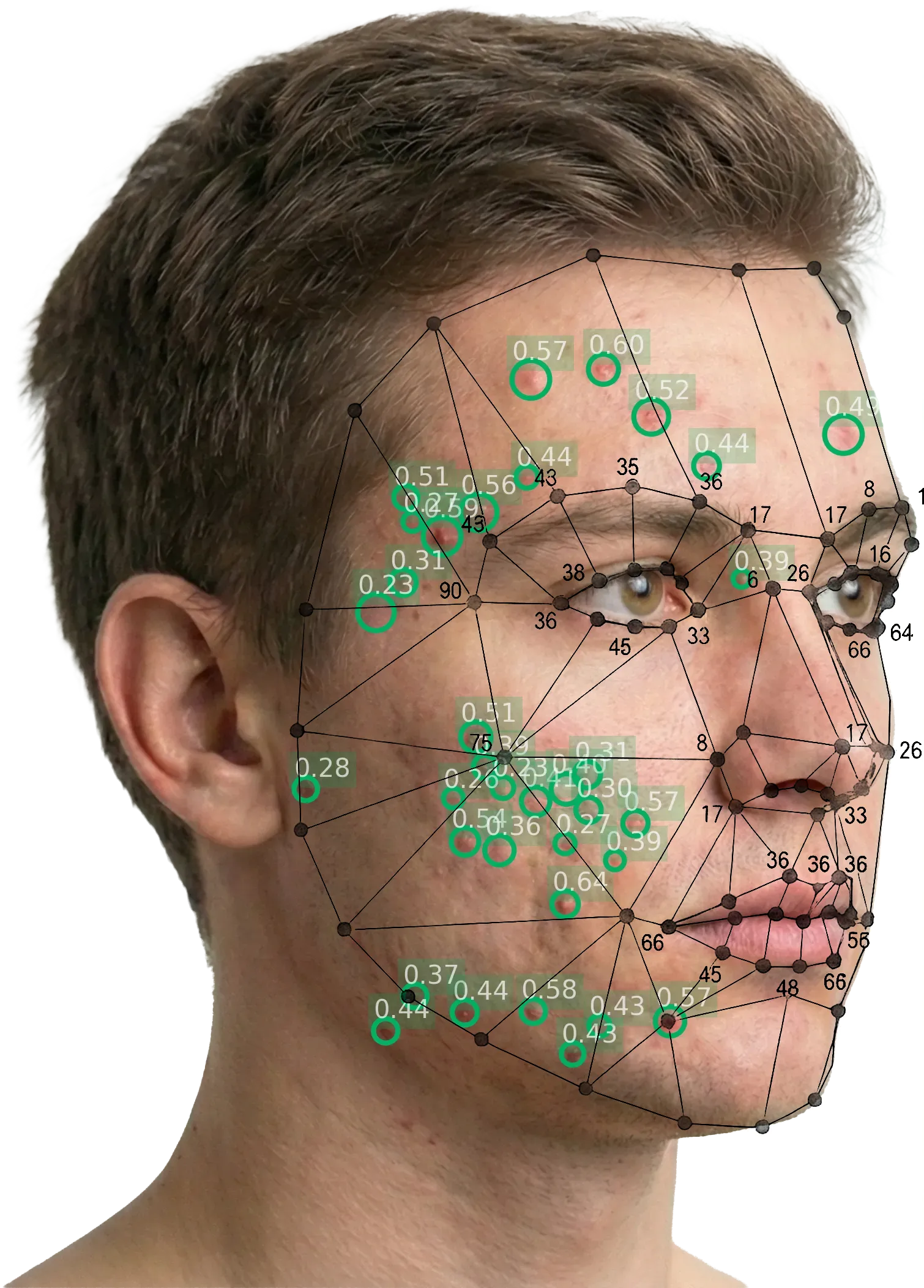
Facial landmark mesh identifying anatomical reference points and region boundaries
Anonymization
All photographs are processed with automatic face anonymization. The system detects facial features and applies irreversible blurring to ensure no patient is identifiable from the stored or exported images.
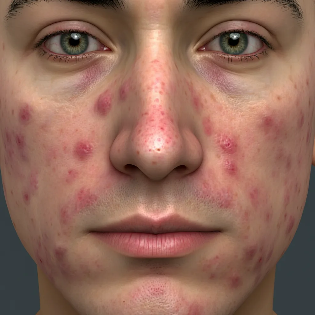
Original capture (eyes, nose, mouth visible)
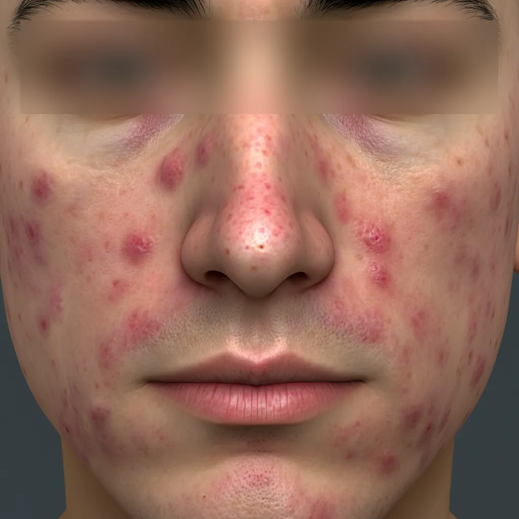
Irreversible face blurring applied
Reliability of the scores
The ALADIN scoring system achieves Cohen's κ = 0.53 against the dermatologist consensus — matching or exceeding the typical inter-rater agreement of individual expert dermatologists (κ = 0.46). This means the AI is at least as consistent a rater as adding a board-certified dermatologist to the panel.
Critically, unlike a human rater, the AI produces the identical score for the same image every time, across every site, without calibration drift or fatigue. There is zero intra-rater variability.
For the full validation evidence: Clinical Evidence →