Imaging Protocol
This page describes the image capture methodology, quality control system, and protocol flexibility available for acne clinical trials using Legit.Health.
Smartphone-based capture
Legit.Health uses standard smartphone cameras for image acquisition. No specialised photography equipment is required.
Traditional clinical photography often relies on systems like Canfield VISIA, which require per-site hardware, per-site calibration, and significant rental or purchase costs. Smartphone-based capture eliminates these costs while maintaining the image quality needed for AI scoring.
The Legit.Health mobile application guides investigators through the capture process with visual perspective silhouettes, real-time DIQA quality checks, and immediate feedback on image adequacy.
Standard 2-perspective protocol
The default imaging protocol follows the Hayashi Criterion (Hayashi et al., 2008), which assesses acne severity by counting inflammatory lesions per half-face. Two photographs are captured at approximately 70-degree diagonal angles:


This protocol ensures that the majority of the facial acne area is visible across the two perspectives, with minimal overlap between them.

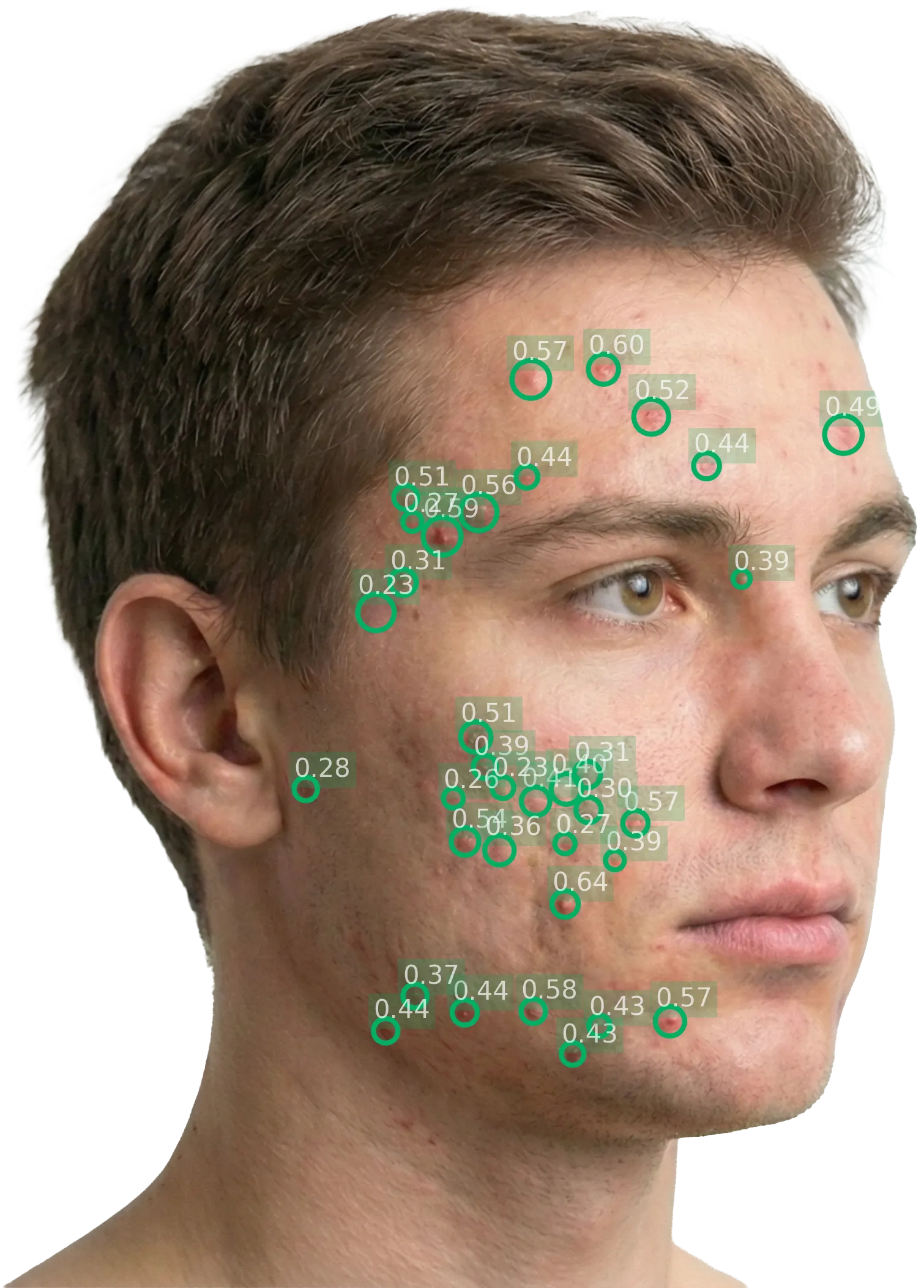
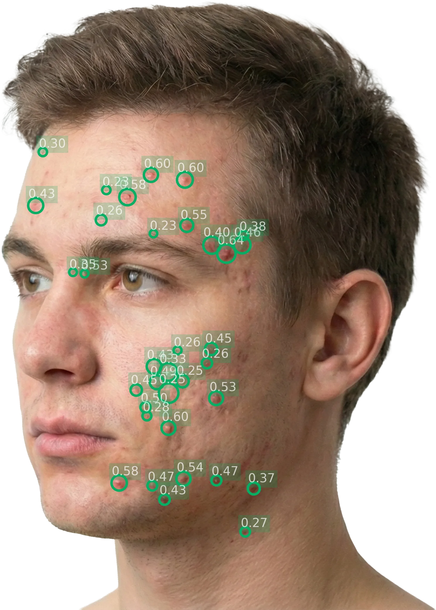
After AI processing, each perspective is annotated with bounding boxes around detected inflammatory lesions:
Alternative perspective protocols
The 2-perspective diagonal protocol is the default, but Legit.Health supports configurable alternatives to match any study protocol:
| Protocol | Perspectives | Use case |
|---|---|---|
| Standard (Hayashi Criterion) | 2 views: Left diagonal (~70°), Right diagonal (~70°) | Most acne studies; captures majority of facial acne area per the Hayashi Criterion for counting inflammatory lesions per half-face. |
| Three-perspective | 3 views: Left perpendicular, Frontal, Right perpendicular | Studies requiring full-face frontal coverage. The frontal view overlaps with both lateral views; facial landmark detection deduplicates lesions. |
| Custom | Any combination of perspectives | Any combination of perspectives, defined in collaboration with the sponsor during protocol design. |
DIQA: Dermatology Image Quality Assessment
What is DIQA?
DIQA (Dermatology Image Quality Assessment) is an AI-powered image quality assessment algorithm that evaluates every captured image in real time before it is accepted for analysis. It was developed by Legit.Health and published in the Journal of the American Academy of Dermatology (Hernández Montilla et al., 2023).
What DIQA evaluates
| Quality dimension | What it checks | Why it matters |
|---|---|---|
| Focus | Sharpness of the image; absence of motion blur | Out-of-focus images can obscure small lesions, leading to undercounting |
| Lighting | Adequate, even illumination; absence of harsh shadows or glare | Poor lighting creates shadows that mimic or hide lesions |
| Framing | Correct anatomical region captured at the required angle | Incorrect framing means the AI analyses the wrong area |
| Resolution | Sufficient pixel density for lesion detection | Low resolution makes small features undetectable |
How it works in the workflow
- The investigator captures an image through the mobile application
- DIQA evaluates the image immediately (sub-second processing)
- If the image passes: it is accepted and queued for AI scoring
- If the image fails: the investigator receives immediate feedback explaining the quality issue and must recapture
Configurable thresholds
The DIQA pass/fail threshold is configurable per study protocol. Sponsors can choose stricter thresholds for pivotal studies (rejecting more images to ensure the highest quality) or more lenient thresholds for real-world evidence studies.
Site instructions and standardisation
Patient preparation
- Remove makeup: Foundation, concealer, and other cosmetics can cover acne lesions
- Pull hair back: Hair should not cover the forehead, cheeks, or jawline
- Remove glasses: Glasses create reflections and shadows that interfere with detection
- Keep facial hair consistent: Facial hair should be maintained consistently throughout the study to minimise variability between visits
Environmental conditions
- Patient positioning: Seated comfortably, remaining still during capture
- Background: Neutral, non-reflective background to reduce artefacts
- Lighting: Well-lit environment with even illumination. Natural light or the smartphone flash can be used. Avoid harsh directional lighting that creates deep shadows.
Consistency across visits
The most important principle is consistency: the same lighting conditions, the same distance from camera to face (approximately 30–40 cm), the same angles, and the same patient preparation at every visit. Consistent capture conditions ensure that score changes between visits reflect actual clinical changes, not variations in image acquisition.
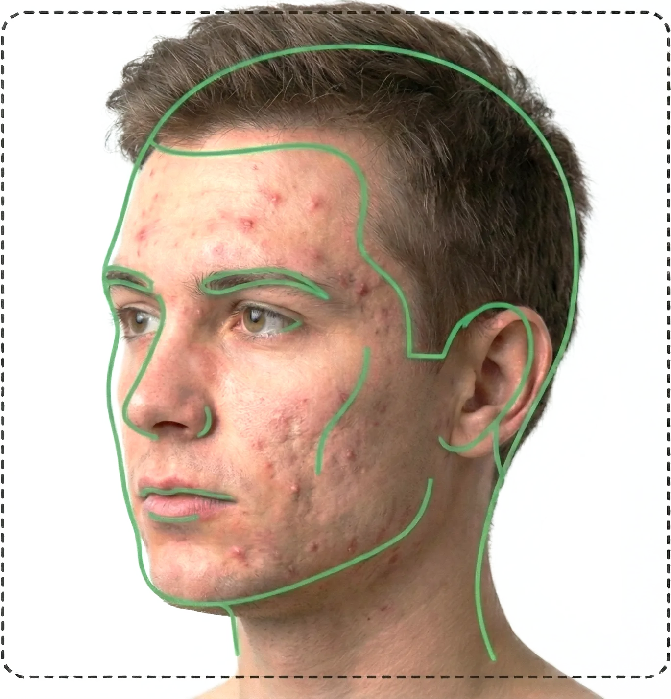
Facial landmark detection
How it works
The AI includes a facial landmark detection module that identifies anatomical reference points in each captured image: eyes, nose, mouth, jawline, and forehead boundaries. The landmark mesh maps the face into regions, allowing the system to determine which lesions belong to each perspective and which should be excluded.
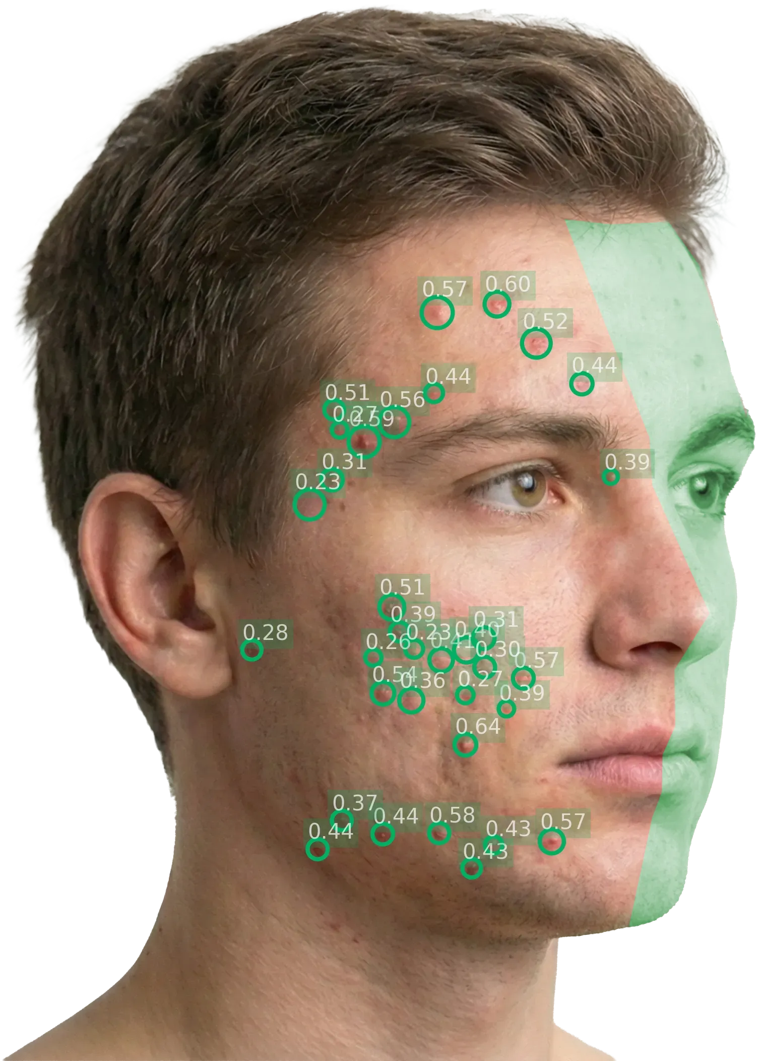
Left diagonal: the green overlay marks the region excluded from this perspective's lesion count. Only lesions on the visible (left) half-face are scored.
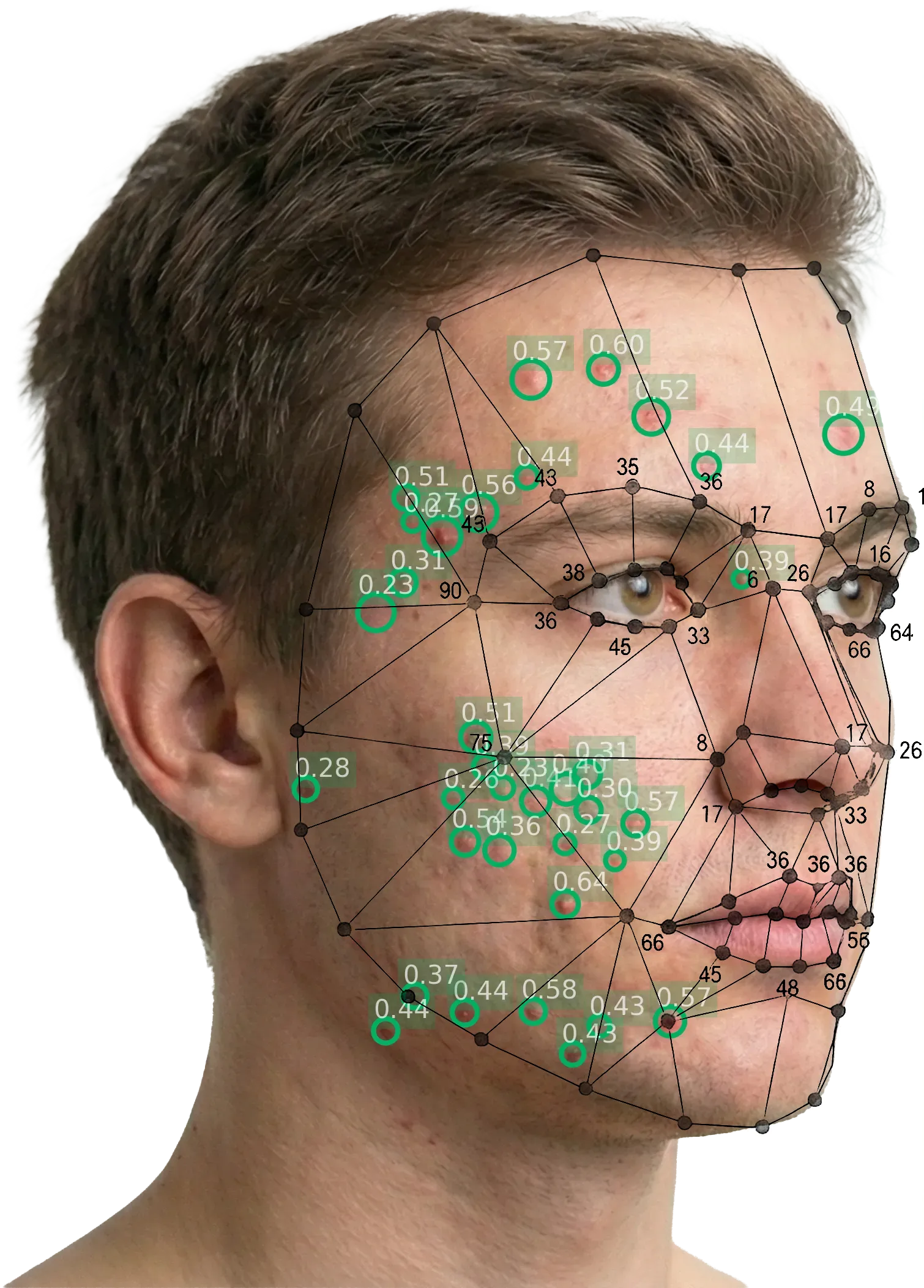
The facial landmark mesh identifies anatomical reference points and defines region boundaries, enabling the system to attribute each detected lesion to a specific half-face.
Applications in the scoring pipeline
Facial landmarks serve multiple purposes:
- Perspective identification: The system confirms which angle the image was captured from by analysing the spatial relationship between landmarks
- Region mapping: Each perspective is mapped to specific facial regions (left cheek, right cheek, forehead, chin, etc.)
- Overlap calculation: When two perspectives cover overlapping facial regions, the landmarks identify the overlap zone
- Lesion deduplication: Lesions detected in the overlap zone of two perspectives are counted only once, preventing inflation of the total count
- Face segmentation: The landmarks define the boundary of the face, ensuring that the AI only analyses the facial area and ignores background elements
Enabling flexible protocols
Facial landmark detection is what makes multi-perspective protocols possible without sacrificing count accuracy. In a 3-perspective protocol (left, frontal, right), the frontal image overlaps significantly with both lateral images. Without landmark-based deduplication, lesion counts from three perspectives would be inflated by double-counted lesions in the overlap zones. The landmark AI solves this by attributing each lesion to a specific facial region and ensuring it is counted exactly once, regardless of how many perspectives capture it.