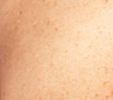
Imaging Protocol
This page describes the image capture methodology, quality control system, and protocol flexibility available for atopic dermatitis clinical trials using Legit.Health.
Smartphone-based capture
Legit.Health uses standard smartphone cameras for image acquisition. No specialised photography equipment is required.
Traditional clinical photography often relies on systems like Canfield VISIA, which require per-site hardware, per-site calibration, and significant rental or purchase costs. Smartphone-based capture eliminates these costs while maintaining the image quality needed for AI scoring.
The Legit.Health mobile application guides investigators through the capture process with visual perspective silhouettes, real-time DIQA quality checks, and immediate feedback on image adequacy.
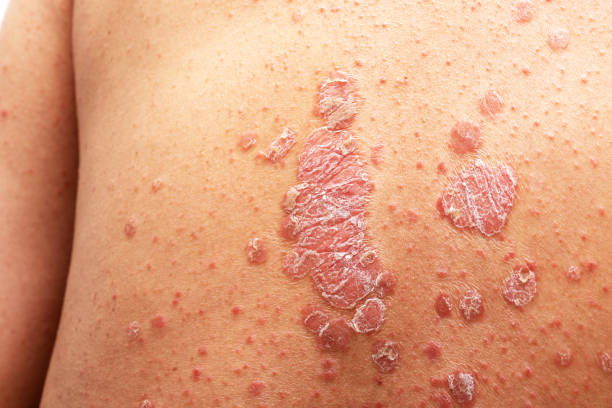
Standard multi-body-site protocol
The default protocol captures 8 images: 4 full-body perspectives for BSA segmentation and 4 close-ups for intensity sign scoring. The app guides the investigator or patient through each perspective in order, with silhouette guidance and a real-time quality check before moving to the next.
8 images (4 perspectives + 4 close-ups)
Perspectives
 ✓
✓ ✓
✓ ✓
✓ ✓
✓Close-ups
 ✓
✓ ✓
✓ ✓
✓ ✓
✓| Body region | Weight | Erythema | Edema | Excoriation | Lichenification | BSA % | Area score | Region EASI |
|---|---|---|---|---|---|---|---|---|
| Head | 0.1 | 0 | 0 | 0 | 0 | 15% | 2 | 0.0 |
| Trunk | 0.3 | 2 | 2 | 2 | 1 | 20% | 2 | 4.2 |
| Upper extremities | 0.2 | 2 | 1 | 2 | 1 | 15% | 2 | 2.4 |
| Lower extremities | 0.4 | 3 | 2 | 2 | 2 | 20% | 2 | 7.2 |
Eczema Area and Severity Index
EASI Score: 13.8
Capture time
The full 8-perspective protocol takes approximately 3–5 minutes using the guided Legit.Health mobile application.
Methods and statistical analysis for EASI
Alternative perspective protocols
The 8-perspective protocol is the default, but the body area coverage can be adapted to match any study design:
| Protocol | Perspectives | Use case |
|---|---|---|
| Full body (8) | 4 body + 4 close-ups | Complete SCORAD/EASI with BSA |
| Flexural focus | 4 perspectives (antecubital + popliteal) | Flexural-dominant AD protocols |
| Face only | 1–3 perspectives | Facial AD studies |
| Target lesion | 1–2 close-ups | Intensity scoring only (no BSA) |
Local EASI from visual signs
A full EASI assessment requires a standardised set of images. When part of that set is missing, the platform degrades gracefully and falls back to a Local EASI: visual signs are scored from whichever close-ups are available, even when BSA-dependent inputs are absent.
Each row below pairs the required input for EASI (left) with a partial dataset (right) showing how visual signs are still computed when imagery is missing.
8 images (4 perspectives + 4 close-ups)
Perspectives
✓✓✓✓Close-ups
✓✓✓✓| Body region | Weight | Erythema | Edema | Excoriation | Lichenification | BSA % | Area score | Region EASI |
|---|---|---|---|---|---|---|---|---|
| Head | 0.1 | 0 | 0 | 0 | 0 | 15% | 2 | 0.0 |
| Trunk | 0.3 | 2 | 2 | 2 | 1 | 20% | 2 | 4.2 |
| Upper extremities | 0.2 | 2 | 1 | 2 | 1 | 15% | 2 | 2.4 |
| Lower extremities | 0.4 | 3 | 2 | 2 | 2 | 20% | 2 | 7.2 |
Eczema Area and Severity Index
EASI Score: 13.8
5 images (4 perspectives + 1 close-up)
Perspectives
✓✓✓✓Close-ups
✗✓✗✗| Body region | Weight | Erythema | Edema | Excoriation | Lichenification | BSA % | Area score | Region EASI |
|---|---|---|---|---|---|---|---|---|
| Head | 0.1 | - | - | - | - | 15% | 2 | - |
| Trunk | 0.3 | 2 | 2 | 2 | 1 | 20% | 2 | 4.2 |
| Upper extremities | 0.2 | - | - | - | - | 15% | 2 | - |
| Lower extremities | 0.4 | - | - | - | - | 20% | 2 | - |
Eczema Area and Severity Index
EASI Score: -
EASI score not available: incomplete dataset (missing regions).
Local EASI and Visual signs available
8 images (4 perspectives + 4 close-ups)
Perspectives
✓✓✓✓Close-ups
✓✓✓✓| Body region | Weight | Erythema | Edema | Excoriation | Lichenification | BSA % | Area score | Region EASI |
|---|---|---|---|---|---|---|---|---|
| Head | 0.1 | 0 | 0 | 0 | 0 | 15% | 2 | 0.0 |
| Trunk | 0.3 | 2 | 2 | 2 | 1 | 20% | 2 | 4.2 |
| Upper extremities | 0.2 | 2 | 1 | 2 | 1 | 15% | 2 | 2.4 |
| Lower extremities | 0.4 | 3 | 2 | 2 | 2 | 20% | 2 | 7.2 |
Eczema Area and Severity Index
EASI Score: 13.8
4 images (0 perspectives + 4 close-ups)
Perspectives
✗✗✗✗Close-ups
✓✓✓✓| Body region | Weight | Erythema | Edema | Excoriation | Lichenification | BSA % | Area score | Region EASI |
|---|---|---|---|---|---|---|---|---|
| Head | 0.1 | 0 | 0 | 0 | 0 | - | - | - |
| Trunk | 0.3 | 2 | 2 | 2 | 1 | - | - | - |
| Upper extremities | 0.2 | 2 | 1 | 2 | 1 | - | - | - |
| Lower extremities | 0.4 | 3 | 2 | 2 | 2 | - | - | - |
Eczema Area and Severity Index
EASI Score: -
EASI score not available: incomplete dataset (missing regions).
Visual signs available
When BSA-required perspectives are missing but representative close-up lesion images are available, EASI visual signs are still scored. AI and clinician panel scores are then compared on the visual-signs dimension only.
Methods for EASI visual signs Analysis
If BSA-required images are missing but representative close-up lesion images are available, EASI visual signs are still scored. AI and clinician panel scores are then compared on the visual-signs dimension only.
4 images (0 perspectives + 4 close-ups)
Representative close-up lesion images
 ✓
✓ ✓
✓ ✓
✓ ✓
✓| Body region | Weight | Erythema | Edema | Excoriation | Lichenification | BSA % | Area score | Region EASI |
|---|---|---|---|---|---|---|---|---|
| Head | 0.1 | 0 | 0 | 0 | 0 | - | - | - |
| Trunk | 0.3 | 2 | 2 | 2 | 1 | - | - | - |
| Upper extremities | 0.2 | 2 | 1 | 2 | 1 | - | - | - |
| Lower extremities | 0.4 | 3 | 2 | 2 | 2 | - | - | - |
Eczema Area and Severity Index
EASI Score: N/A
EASI score not available: incomplete dataset (missing regions).
Visual signs available
4 images (0 perspectives + 4 close-ups)
Representative close-up lesion images
✓✓✓✓| Body region | Weight | Erythema | Edema | Excoriation | Lichenification | BSA % | Area score | Region EASI |
|---|---|---|---|---|---|---|---|---|
| Head | 0.1 | 0 | 0 | 0 | 0 | - | - | - |
| Trunk | 0.3 | 2 | 2 | 2 | 1 | - | - | - |
| Upper extremities | 0.2 | 2 | 1 | 2 | 1 | - | - | - |
| Lower extremities | 0.4 | 3 | 2 | 2 | 2 | - | - | - |
Eczema Area and Severity Index
EASI Score: N/A
EASI score not available: incomplete dataset (missing regions).
Visual signs available
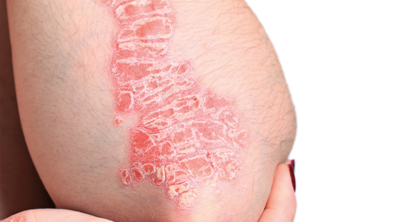
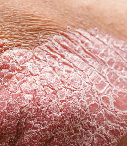
DIQA: Dermatology Image Quality Assessment
What is DIQA?
DIQA (Dermatology Image Quality Assessment) is an AI-powered image quality assessment algorithm that evaluates every captured image in real time before it is accepted for analysis. It was developed by Legit.Health and published in the Journal of the American Academy of Dermatology (Hernández Montilla et al., 2023).
What DIQA evaluates
| Quality dimension | What it checks | Why it matters |
|---|---|---|
| Focus | Sharpness of the image; absence of motion blur | Out-of-focus images can obscure small lesions, leading to undercounting |
| Lighting | Adequate, even illumination; absence of harsh shadows or glare | Poor lighting creates shadows that mimic or hide lesions |
| Framing | Correct anatomical region captured at the required angle | Incorrect framing means the AI analyses the wrong area |
| Resolution | Sufficient pixel density for lesion detection | Low resolution makes small features undetectable |
How it works in the workflow
- The investigator captures an image through the mobile application
- DIQA evaluates the image immediately (sub-second processing)
- If the image passes: it is accepted and queued for AI scoring
- If the image fails: the investigator receives immediate feedback explaining the quality issue and must recapture
Configurable thresholds
The DIQA pass/fail threshold is configurable per study protocol. Sponsors can choose stricter thresholds for pivotal studies (rejecting more images to ensure the highest quality) or more lenient thresholds for real-world evidence studies.
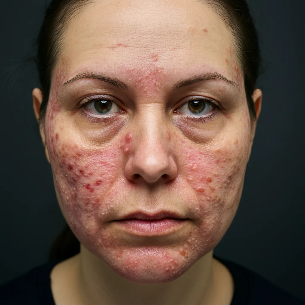
Patient preparation
- Remove clothing from the area being photographed
- No moisturiser or topical treatments applied within 2 hours of the visit (unless protocol specifies otherwise)
- Hair pinned back for face and neck assessments
- Remove jewellery from the area being photographed
- Neutral, well-lit background; standard smartphone flash or even natural light
Environmental conditions
- Patient positioning: Seated or standing comfortably, remaining still during capture
- Background: Neutral, non-reflective background to reduce artefacts
- Lighting: Well-lit environment with even illumination. Natural light or smartphone flash can be used. Avoid harsh directional lighting that creates deep shadows.
- Distance: Approximately 30–50 cm from camera to skin surface, adjusted per body area
Consistency across visits
The most important principle is consistency: the same lighting conditions, the same distance, the same angles, and the same patient preparation at every visit. Consistent capture conditions ensure that score changes between visits reflect actual clinical changes, not variations in image acquisition.
Anonymization
All photographs are processed with automatic face anonymization. The system applies irreversible blurring to facial features, ensuring patient privacy for stored and exported images.
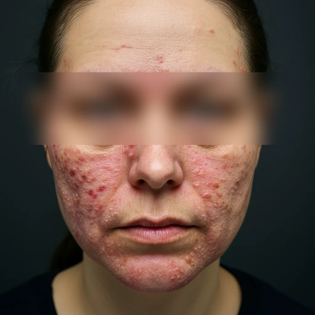
Irreversible face blurring applied