Cutaneous Lupus Erythematosus Severity Endpoints for Clinical Trials
The AI scoring provided by Legit.Health automates the visual components of the CLASI (Cutaneous Lupus Erythematosus Disease Area and Severity Index), the reference outcome measure in cutaneous lupus erythematosus (CLE) drug development. It quantifies the erythema, scale, induration, pigment loss and hair loss that CLASI is built from, from standard smartphone images, and assembles them into the CLASI Activity and Damage scores.
CLASI Activity
CLASI-A 11
Reversible activity, range 0–70
CLASI Damage
CLASI-D 6
Cumulative damage, range 0–56
Timestamp
10/2/2026, 1:27:30 AM
Analysis performed in
1.6 seconds
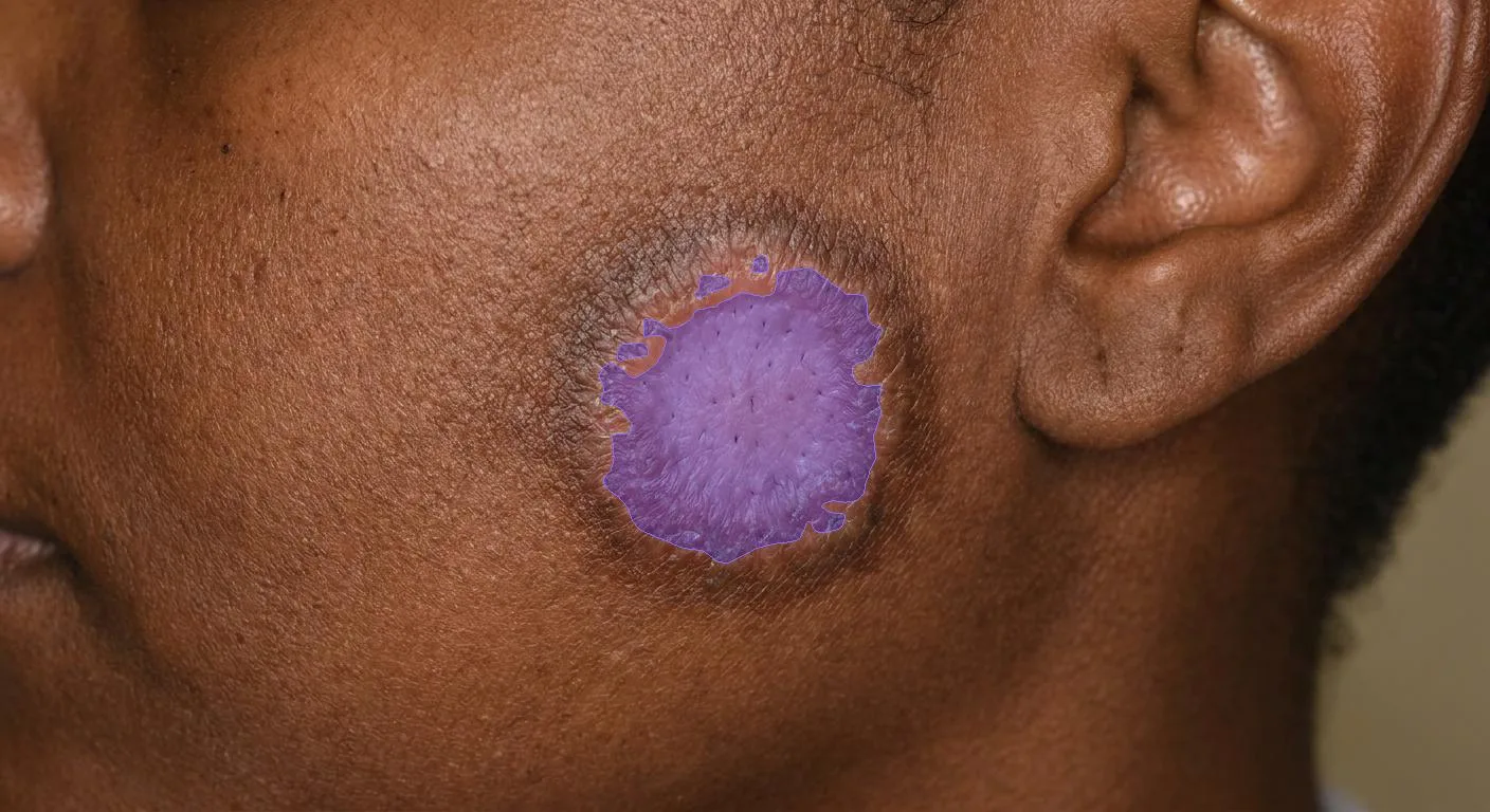
Region
Nose and malar area
Image quality (DIQA)
83%
Local CLASI-A (E + S)
3
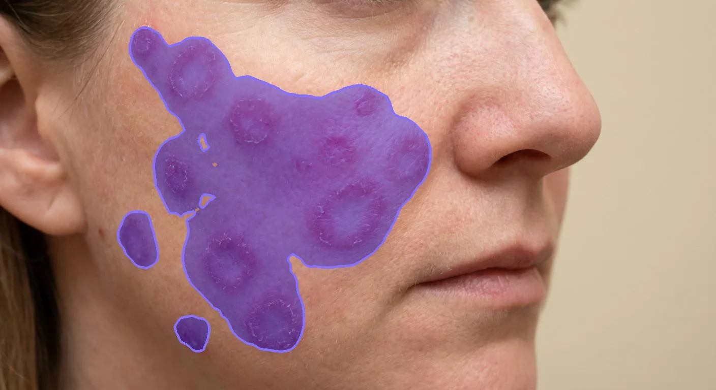
Region
Rest of face
Image quality (DIQA)
91%
Local CLASI-A (E + S)
4
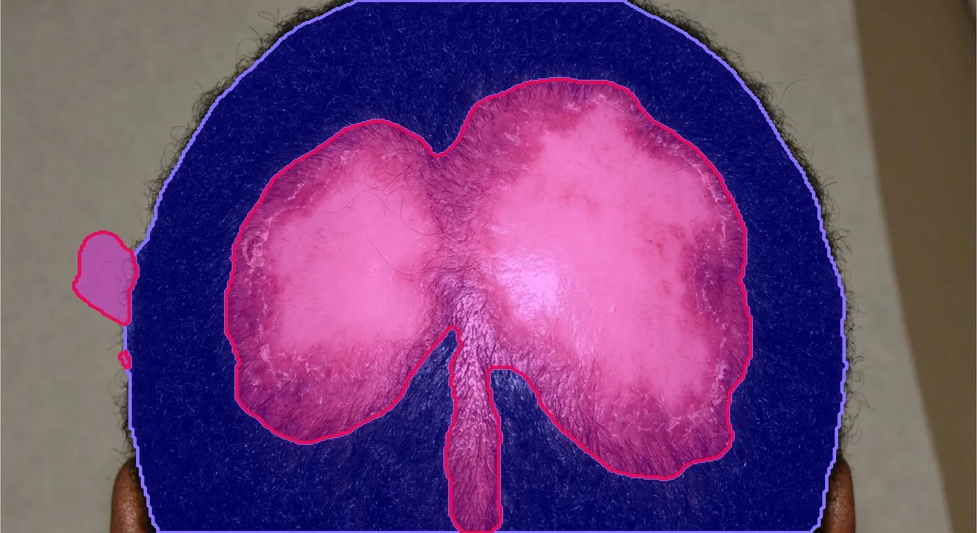
Region
Scalp
Image quality (DIQA)
81%
Local CLASI-A (E + S)
1
Example per-visit CLASI report. Synthetic imagery, real device outputs for the signs shown; the full report set is on the Sample outputs page.
The CLASI instrument
CLASI is the validated reference instrument for CLE, developed by Albrecht and colleagues (2005) and adopted as the outcome measure in cutaneous lupus trials. It is unusual among dermatological indices because it produces two independent scores from one examination:
Erythema and scale/hypertrophy per region, plus alopecia and mucosal involvement
Responds to treatment within weeks; the usual efficacy endpoint
Dyspigmentation and scarring/atrophy per region, plus scalp scarring
Accumulates irreversibly; the long-term burden measure
Both scores come from the same set of photographs, and separating activity from damage reliably is what makes each one interpretable. The instrument's own validation and the literature behind it are on the Clinical evidence and Publications pages.
Endpoint capabilities
The platform provides four configurable endpoints, tailored to the study protocol:
| Endpoint | Definition | AI output | Typical use in protocol |
|---|---|---|---|
| CLASI-A (Activity) | Reversible disease activity: erythema and scale/hypertrophy per region, plus alopecia and mucosal involvement | Activity score 0–70, from measured signs plus clinician-entered items | Primary |
| CLASI-D (Damage) | Irreversible damage: dyspigmentation and scarring per region, plus scalp scarring | Damage score 0–56 | Secondary |
| Depigmentation extent | Continuous depigmented area per lesion and region; the basis of a repigmentation measure the binary CLASI-D item cannot express | Affected area, relative or mm² with marker capture | Exploratory |
| Per-sign intensity and extent | Erythema, scale and induration as continuous per-region measures, at higher resolution than the CLASI item scales | Per-sign value and area per region | Exploratory |
CLASI is a coarse instrument by design: erythema is a four-point item, dyspigmentation a single present-or-absent flag. The device measures the same signs continuously, so a study can report the CLASI scores regulators expect and the higher-resolution per-sign measures that detect a treatment effect earlier. The clearest case is repigmentation: a depigmented plaque refilling with pigment is a change patients value, and one CLASI-D structurally cannot show, but a continuous depigmentation extent can.
What the AI measures
Each measurement is produced from one photograph of one anatomical region.
| Measurement | Output | CLASI component it serves |
|---|---|---|
| Erythema intensity | Graded severity of redness | CLASI-A erythema |
| Erythema extent | Affected area, relative or absolute | Lesion burden within the region |
| Desquamation intensity | Graded severity of scaling | CLASI-A scale |
| Induration intensity | Graded plaque thickening | CLASI-A hypertrophy |
| Depigmentation extent | Affected area, relative or absolute | CLASI-D dyspigmentation |
| Lesion area | Physical area in mm², with marker capture | Longitudinal lesion measurement |
| Hair loss percentage | Affected proportion of the scalp | CLASI-A and CLASI-D scalp items |
| Diagnosis support | Ranked differential including CLE | Screening and eligibility review |
| DIQA image quality | Quality score with accept or recapture | Applies to every capture |
| Skin and body segmentation | Pixel-level masks | Underlies every extent figure |
The full item-by-item mapping, including the CLASI components that are not derived from an image, is on the Scoring methodology page.
How the AI works
The pipeline runs in three stages, from photograph to assembled score.
Stage 1: Image quality gate
Every image is assessed by DIQA (Dermatology Image Quality Assessment) before any sign is read. Captures whose exposure, focus or framing fall outside the acceptable range are rejected for recapture, so a poor photograph produces a prompt rather than a wrong number. See the Imaging protocol.
Stage 2: Per-sign measurement
Dedicated models measure each visual sign in the photographed region: erythema, desquamation and induration for activity; depigmentation for damage; hair loss on the scalp. Each sign is returned with a confidence figure and a pixel-level segmentation mask.
An extent is reported two ways. Relative extent divides the segmented sign area by the photographed region:
Where a calibration marker of known side (mm) spanning pixels is in frame, the segmented pixel count converts to absolute area in physical units:
The absolute form is independent of working distance, which is what makes it comparable across visits; the imaging protocol covers marker capture. Every sign is also qualified by the scoring system that produced it, because the same sign is scored on different scales by different validated instruments and the values are not interchangeable; see scale alignment.
Stage 3: CLASI assembly
Per-region sign values are summed into the two CLASI scores. The activity score adds the two activity signs across the scored anatomical regions, plus the three clinician-entered items:
with erythema (0–3), scale or hypertrophy (0–2), mucous-membrane involvement (0–1), recent hair loss (0–1) and non-scarring alopecia (0–3); total range 0–70.
The damage score adds dyspigmentation and scarring, with the dyspigmentation subtotal doubled when it persists beyond twelve months:
with dyspigmentation (0–1), the persistence flag , scarring or atrophy (0–2) and scalp scarring ; total range 0–56.
The device measures the imaged terms (, , , and the alopecia extent behind ); the investigator supplies , and the persistence flag at review. Assembly depends on each image being attached to a patient, a visit and a named region; without that, the device returns valid per-image values that cannot be summed. The Imaging protocol sets out the metadata this requires.
CLASI-A severity strata
Activity scores map to severity bands validated by Klein and colleagues (2011):
| CLASI-A score | Severity |
|---|---|
| 0–9 | Mild |
| 10–20 | Moderate |
| 21–70 | Severe |
The same work found that a clinically meaningful response corresponds to roughly a 4-point or 20% fall in the activity score, the basis for a CLASI-A response endpoint. Details and references are on the Clinical evidence page.
Protocol flexibility
The region set, visit structure, target lesions and calibration are configured per study at setup. A study can capture all 13 regions at every visit or take a comprehensive baseline with focused follow-up, and can report absolute lesion area in mm² where calibration markers are used. The Imaging protocol and Trial workflow pages cover the options.