Vitiligo Severity Endpoints for Clinical Trials
The AI scoring provided by Legit.Health delivers automated, standardised vitiligo severity scoring for clinical trials, measuring the extent of depigmented skin in each body region from standardised photographs to compute the Vitiligo Area Scoring Index (VASI), the standard measure of vitiligo extent in clinical trials, together with its facial (F-VASI) and total body (T-VASI) forms.
Legit.Health supports interventional and observational studies across all clinical trial phases, from Phase I to Phase IV, as well as real-world evidence (RWE) and post-marketing studies.
Total Vitiligo Area Scoring Index
0 to 100 scale, the 14 views of the six VASI sites
T-VASI = 1.35 + 12.04 + 5.50 + 3.48 + 9.27 + 2.49 = 34.13
Images analysed
14 (the full capture set)
Analysis performed in
3.4 seconds
- VASI: points the site adds to the T-VASI
- %: depigmentation of the view
- : DIQA image quality
Head and neck
3.5% of the body
VASI1.35
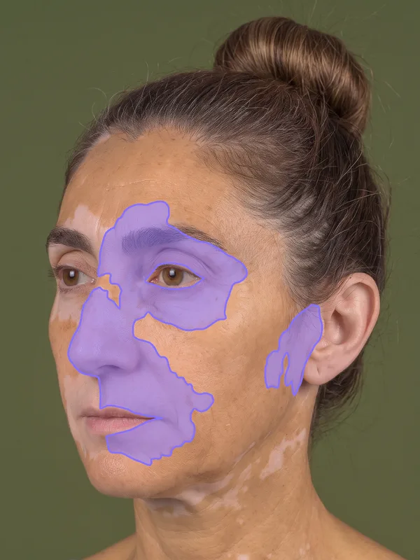
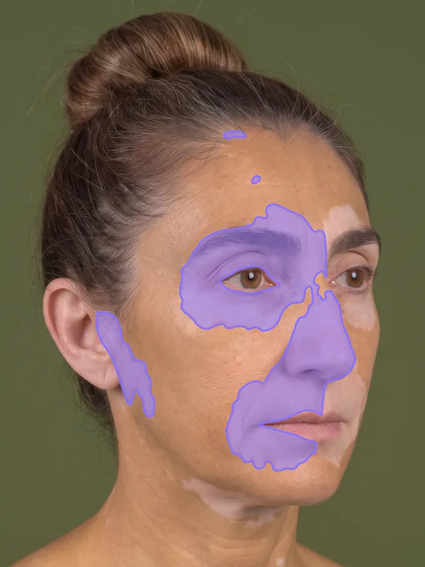
Facial Vitiligo Area Scoring Index
0 to 3.5 scale, the two halves of the face
F-VASI = 0.68 + 0.67 = 1.35
Trunk
28% of the body
VASI12.04
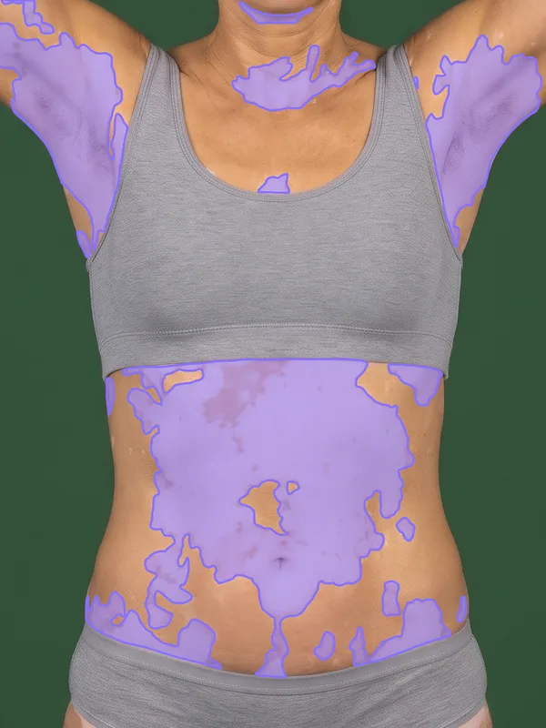
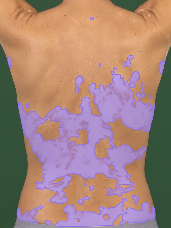
Lower extremities
38% of the body
VASI9.27
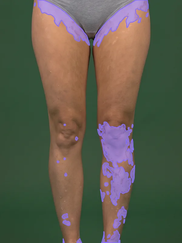
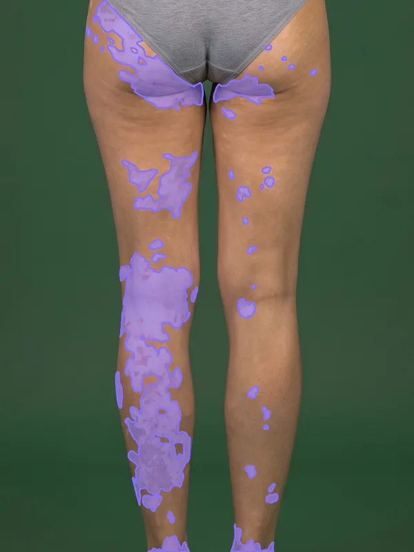
Feet
7% of the body
VASI2.49
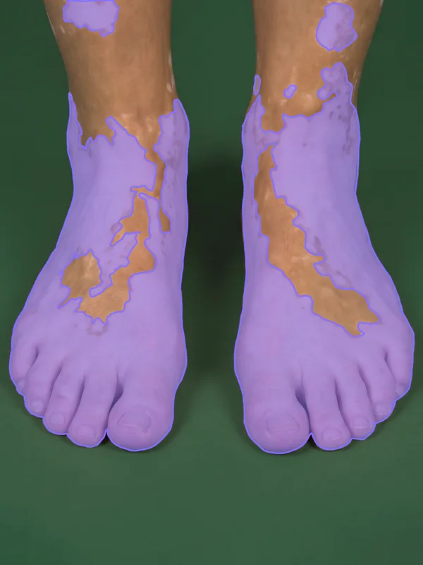
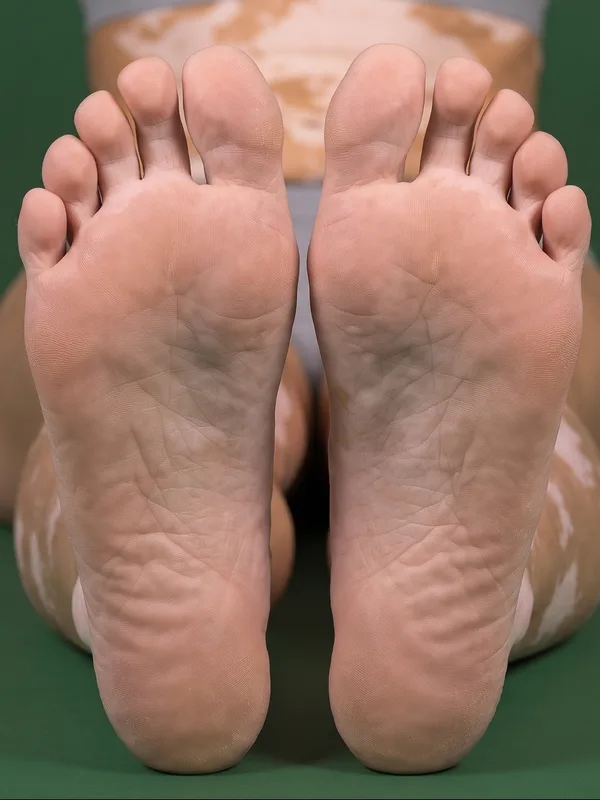
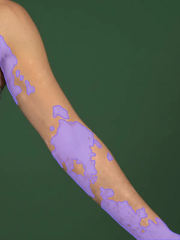
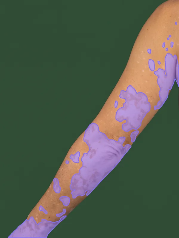
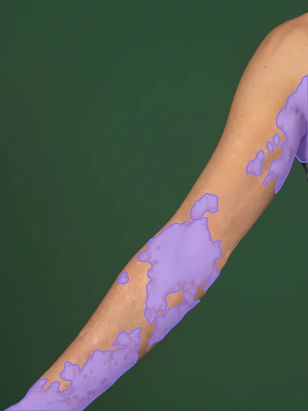
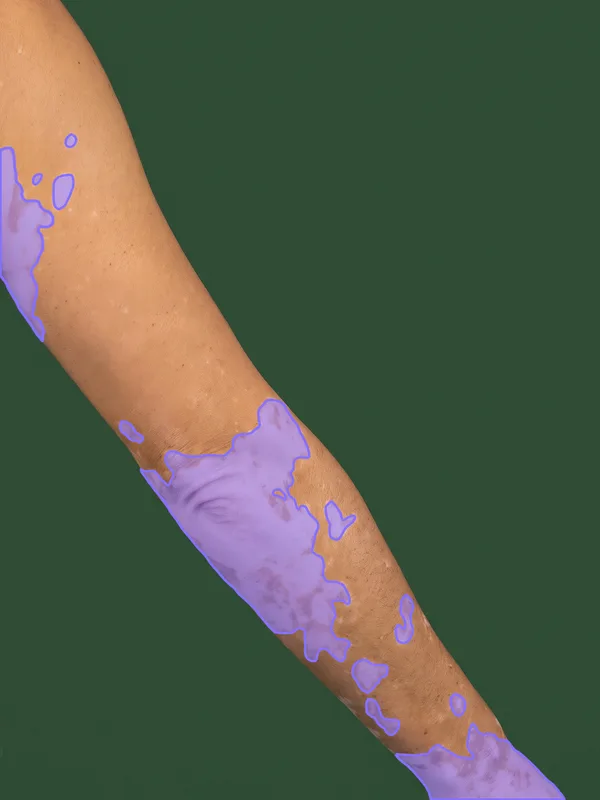
Upper extremities
14% of the body
VASI5.50
Hands
5% of the body
VASI3.48
The validation of the model behind these scores is on Clinical evidence.
The example images on this page are synthetic dermatology imagery and vector illustrations; they are not patient records.
Why automated VASI scoring for clinical trials?
Manual VASI asks a rater to estimate, region by region, how much of the body surface is affected, using the palm of the patient's hand as a unit, and then to grade by eye how much pigment remains in each affected area. Both judgements are operator-dependent, they are made in coarse steps, and a full-body assessment takes a significant share of the visit. Two raters looking at the same patient often land on different values, which adds noise to the change from baseline that vitiligo trials rely on.
The AI measures the depigmented surface the score is built on:
- Depigmentation measurement: pixel-level segmentation of depigmented skin in each photograph, replacing visual estimation with palm units
- VASI calculation: the percentage measured in each view, weighted by the share of the body surface the view represents and added up into the total score
- DIQA (Dermatology Image Quality Assessment): image quality assessed before scoring, so substandard images are flagged for recapture at the visit rather than surfacing as missing data at database lock
The photographs follow a standardised capture protocol, so every visit is photographed the same way; see Imaging protocol.
Automated VASI returns a continuous measurement from standardised photographs, with three properties manual scoring cannot offer:
- Perfect reproducibility: the same image always produces the same score, with zero intra-rater variability, at every site and every visit.
- No investigator training drift: performance does not depend on how recently a site was trained or on which rater is on shift that day.
- Less noise in VASI change from baseline, which can support smaller sample sizes or greater statistical power to detect treatment response, once agreement with investigator scoring is confirmed.
Endpoint capabilities
The AI reports the VASI and its regional components from a single capture set, configurable per study protocol:
- T-VASI
- 0 to 100
- Total body: the percentage of the body surface with depigmentation
- F-VASI
- Face only
- Facial: the same measurement on the two face views, one for each half
- Structure
- 6 body sites
- Head and neck, hands, upper extremities, trunk, lower extremities and feet, as the VASI defines them
- Regional extent
- 0 to 100% per view
- Depigmentation in each view, measured at pixel level, not estimated
VASI is the efficacy measure of vitiligo programmes, and responder definitions such as F-VASI75 and T-VASI50 are read from the same score; see the worked example. The values per view and per site are reported alongside, because they show which part of the body drives the repigmentation.
How the AI works
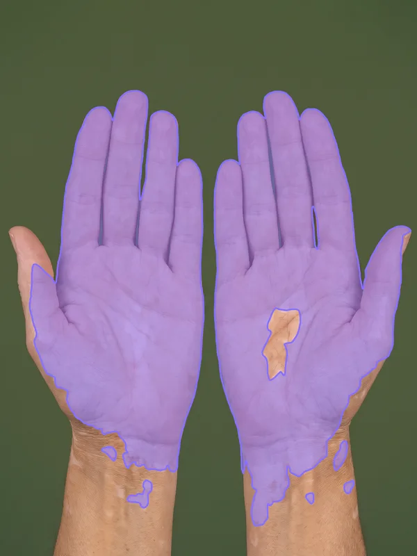
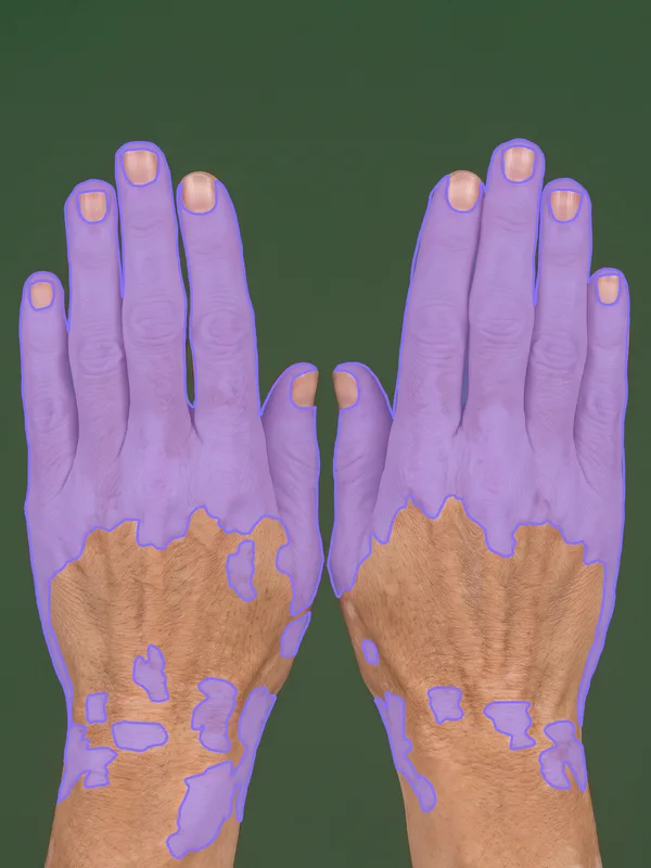
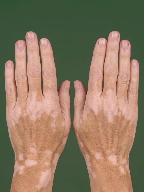
Each photograph shows one side of one body site. For each photograph, a deep learning model separates the skin from the background and classifies every pixel of skin as pigmented or depigmented. It then measures the percentage of the skin affected in that view, and each view, weighted by its share of the body surface, is added into the VASI score.

- Skin, separated from the background

- Depigmented skin
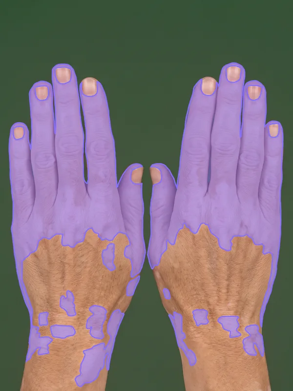
- Result over the photograph
The segmentation mask appears over the original photograph, so the investigator sees exactly which areas were counted as depigmented:
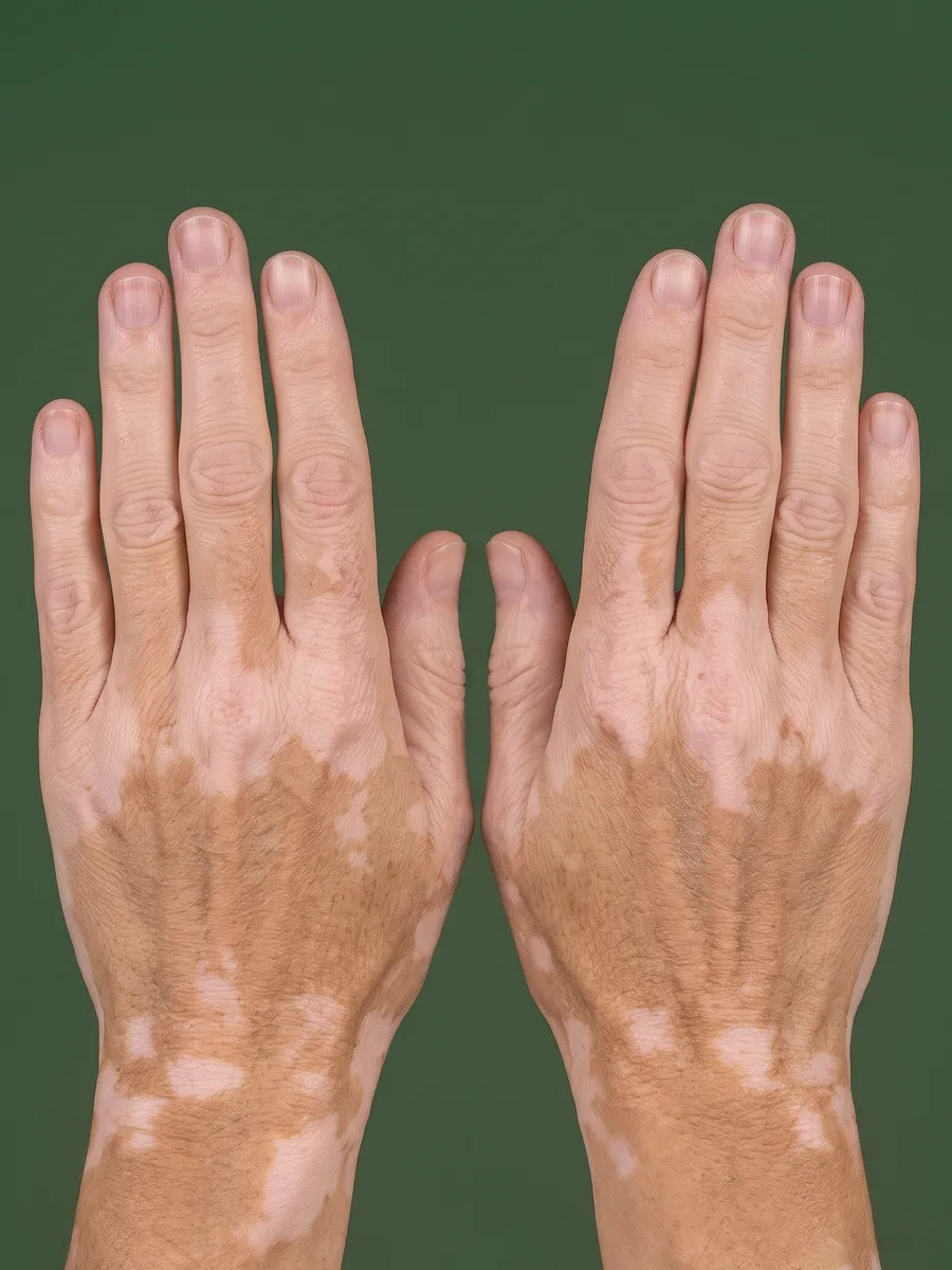
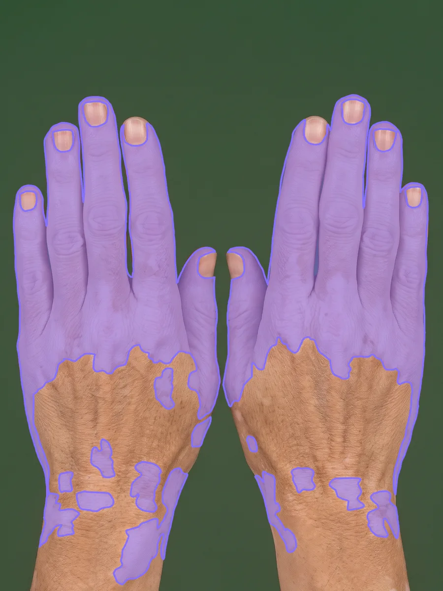
Automated analysis of the backs of the hands: drag the handle to show or hide the depigmented skin the model found. Synthetic image, not a patient record.
Longitudinal severity tracking
From the follow-up visits onward, the same report shows how severity evolves across the study:
- VASI trajectory: absolute and percentage change from baseline at each visit, from screening to end of study
- Responder definitions: whether the patient has reached F-VASI50, F-VASI75, F-VASI90, F-VASI100, T-VASI50 or T-VASI75, derived from the per-visit scores
- Per-region trends: which part of the body is driving repigmentation or further spread
- Alerts against baseline: a notification to the site when depigmentation increases by the configured threshold
- Protocol adherence: whether assessments are captured at the correct intervals
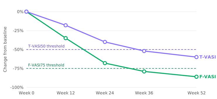
One illustrative patient: F-VASI crosses the F-VASI75 threshold (dashed, lower) between weeks 24 and 36, and T-VASI crosses the T-VASI50 threshold (dashed, upper) by week 36. Illustrative values
The platform provides automated severity scoring as decision support. Scores are interpreted by qualified healthcare professionals within the patient's overall clinical context; the platform does not replace clinical judgement or make autonomous diagnostic decisions.