Scoring Methodology
The platform from Legit.Health scores vitiligo by measuring depigmentation rather than estimating it: the software segments every pixel of skin in each photograph, measures the percentage of depigmented skin in each view and adds up the views into the VASI score, and the investigator reviews the report at the visit. The regional values are reported in their own right, so any responder definition the protocol needs comes from the same capture.
The example images on this page are synthetic dermatology imagery and vector illustrations; they are not patient records.
Depigmentation segmentation
- Photograph: The standard views of the body and the face.
- Separate: Skin from background.
- Segment: Pigmented or depigmented skin per pixel.
- Measure: Depigmentation % per view.
- Score: Each view weighted by its share of the body surface, added up into the facial and total VASI.
A deep learning segmentation model, trained on images annotated by several dermatologists and unified into a consensus reference, assigns every pixel of each photograph to one of three classes.
Normal skin
Skin with its normal pigmentation.
Depigmented skin
Skin with reduced or absent pigmentation: the vitiligo lesions.
Background
Everything that is not skin: clothing, hair and background. Excluded from the calculation.
From photograph to percentage
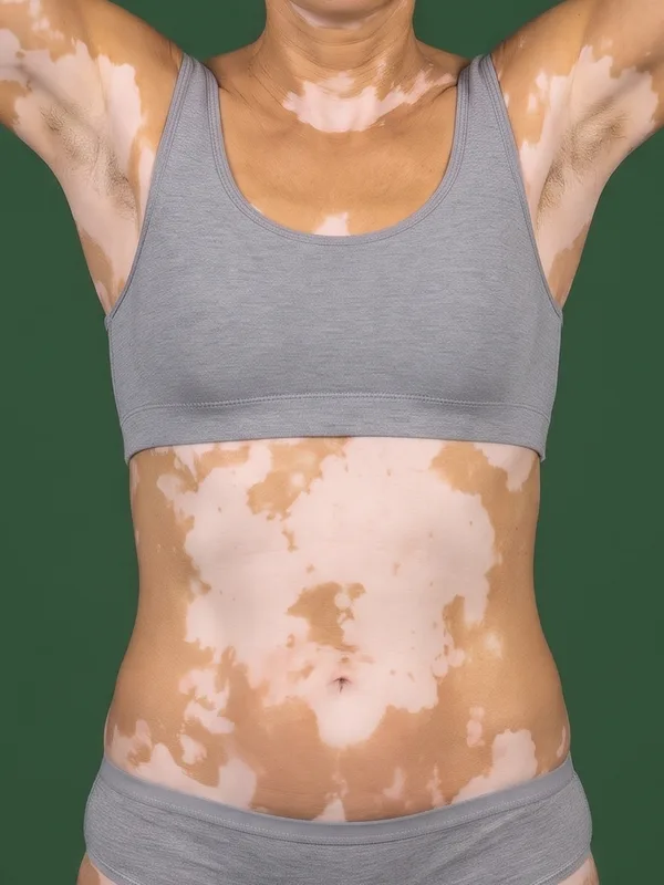
- Photograph of the view
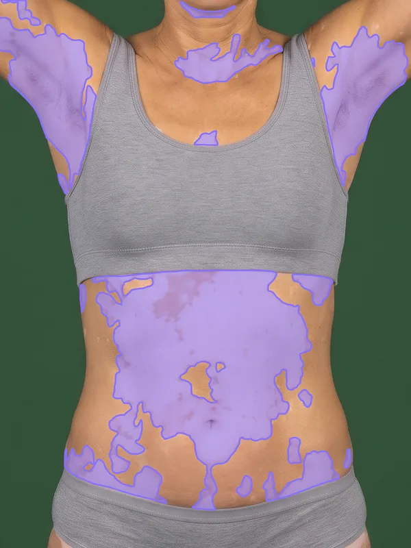
- Depigmented skin found by the model, in violet
- Depigmentation of the view
For each view , the depigmentation percentage is the share of depigmented pixels within the skin that the photograph shows:
Background pixels are left out of both terms, so clothing and the room never count as depigmentation.
VASI
The Vitiligo Area Scoring Index (VASI) is the standard measure of vitiligo extent in clinical trials. The T-VASI scores the whole body, on a scale from 0 to 100, and the F-VASI scores the face alone, from 0 to 3.5, because the face is about 3.5% of the body surface. Both come from the same model and the same photographs.
The six body sites
For the T-VASI, the body is divided into six mutually exclusive sites. Select a site to see what it includes.
The face, the scalp and the neck, front and back.
A large site such as the trunk can contribute more points than the hands only because there is more skin in it. The imaging protocol photographs each site in one or more views, and every view stands for a fixed share of the body surface.
How the automated score relates to the VASI
At the visit, a rater estimates two things for each area of vitiligo: its extent, in hand units of about 1% of the body, and its degree of depigmentation, in seven steps. Multiplying them gives the amount of skin that has lost its pigment: an area of 2 hand units at 50% is 1 hand unit of depigmented skin. Added over the body, the VASI is the depigmented surface of the body, in percent of the body surface.
The photographs measure that surface directly. Each photograph gives two numbers, the pixels of skin and the pixels of depigmented skin. Their ratio is the share of the view's skin that has lost its pigment, and the share of the body surface that the view stands for turns it into body surface:
Adding the views of a site gives that site's points; adding the two face views gives the F-VASI, and adding all the views gives the T-VASI. Counting depigmented pixels measures the product of the extent and the degree at once, so the automated score needs no estimate of either.
For example, a lesion on the chest covers 3 hand units and three quarters of it is still white. A rater scores it as points. The photograph of the front of the trunk, which stands for 14% of the body, shows 168,000 pixels of skin, 27,000 of them depigmented:
- What the model outputs: 27,000 pixels of depigmented skin and 141,000 pixels of normal skin.
- The ratio: of the skin in the photograph is depigmented.
- The points: points, the same as the rater's.
The pixel counts change with the camera and the distance; the ratio, and so the points, do not, so the protocol needs no ruler or calibration marker.
The result is the same information as a manual VASI, on the same scale, so F-VASI75, T-VASI50 and every other responder definition are read from it unchanged. What changes is how it is obtained:
- Measured, not estimated: every pixel of the photograph is counted, instead of the rater's eye judging the area in hand units.
- Continuous: the score is not rounded to seven degrees of depigmentation or to tenths of a hand unit, so smaller changes between visits show.
- Reproducible: the same photographs always give the same score, at every site and every visit.
- Traceable: every point of the score comes from a photograph and its segmentation mask, which can be reviewed afterwards.
The automated score does not report a degree of depigmentation. Where a protocol needs one for each lesion, it is the share of the lesion's outline that has no pigment, rounded to the nearest of the seven steps; deriving it needs the outline of each lesion as well as its depigmented pixels, and it is not part of the current scoring. Where the automated and the manual score can differ in practice is set out in Limitations.
The face
The F-VASI is measured on two views, one for each half of the face, and only the facial area of the F-VASI counts:
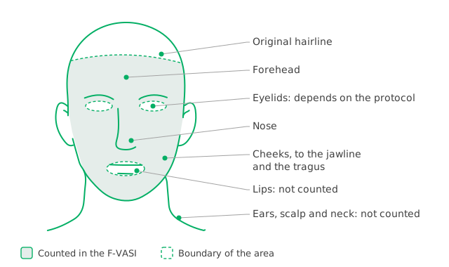
The facial area of the F-VASI: forehead to the original hairline, the cheeks down to the jawline and from the corner of the mouth to the tragus, and the nose. Scalp, lips, ears and neck are left out; some protocols count the eyelids and others exclude them
Overlap between views
The two face views meet in the middle of the face, around the centre of the forehead, the nose and the chin, where the same patch of depigmentation can appear in both photographs. Because the F-VASI adds the two halves rather than averaging them, a patch counted in both would be counted twice. Each view therefore measures only the half of the face that faces the camera: facial landmarks find the midline, and the skin beyond it is left to the other view.
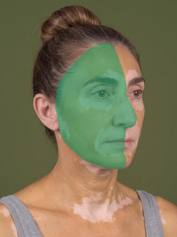
Right view: the green area is the half of the face it measures, up to the midline.
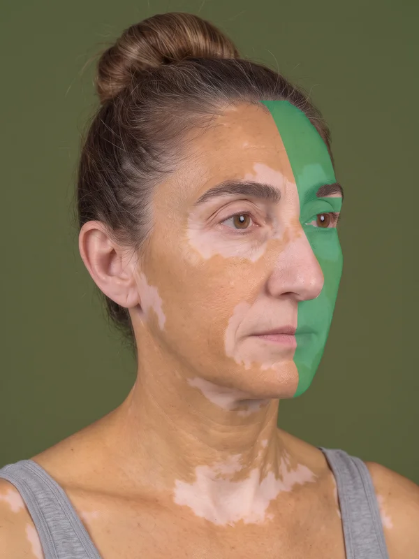
The same view: the green area is the half that the left view measures, so it is not counted here.
A worked example
An illustrative example, not study data: the same patient followed across a 52-week trial, with assessments at baseline and at weeks 12, 24 and 52. The depigmentation of each site is measured on its views at each visit; the F-VASI comes from the two face views and the T-VASI adds up the six sites, and the thresholds show when the patient reaches each response.
Change from baseline
F-VASI and T-VASI, against the 50% and 75% reductions
T-VASI contribution by site
Points each site adds to the total
| Depigmentation per site | Baseline | Week 12 | Week 24 | Week 52 |
|---|---|---|---|---|
| Head and neck (2 face views, 3.5% of the body) | 39% | 29% | 17% | 9% |
| Trunk (2 views, with the neck, 28% of the body) | 43% | 38% | 30% | 17% |
| Upper extremities (4 views, 14% of the body) | 39% | 36% | 28% | 20% |
| Hands (2 views, 5% of the body) | 70% | 69% | 67% | 63% |
| Lower extremities (2 views, 38% of the body) | 24% | 22% | 16% | 9% |
| Feet (2 views, 7% of the body) | 36% | 35% | 34% | 33% |
| F-VASI | 1.35 | 1.03 (-24%) | 0.61 (-55%) | 0.30 (-78%) |
| T-VASI | 34.13 | 30.96 (-9%) | 24.72 (-28%) | 16.79 (-51%) |
| Responses reached | Reference | None yet | F-VASI50 | F-VASI50F-VASI75T-VASI50 |
The face responds first: the patient reaches F-VASI50 at week 24 and F-VASI75 at week 52, the usual primary endpoint, while F-VASI90 is not yet reached. The body follows more slowly and reaches T-VASI50 at week 52. The trunk drives most of the change in the T-VASI: its two views stand for 28% of the body, so its repigmentation from 43% to 17% removes 7.28 of the 17.34 points the score falls, while the hands and the feet change least. The photographs below show the back of the same patient at the first and last visits.
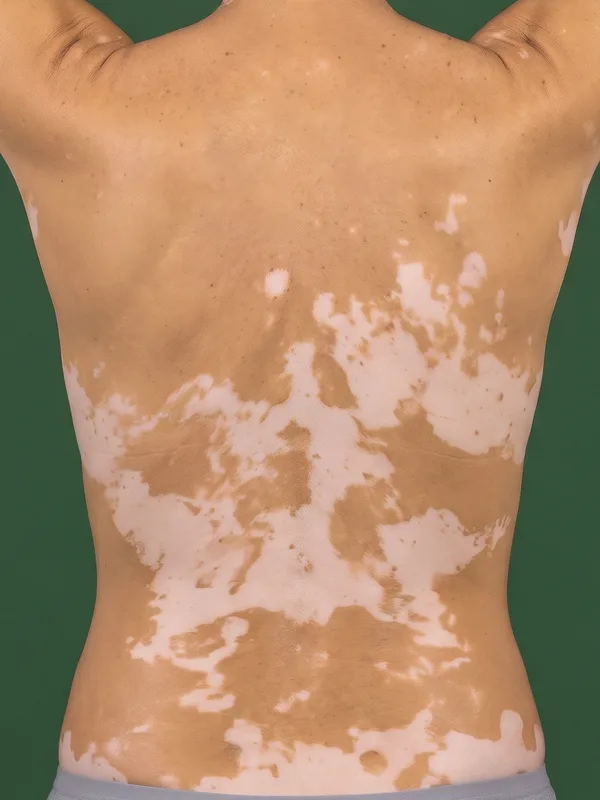
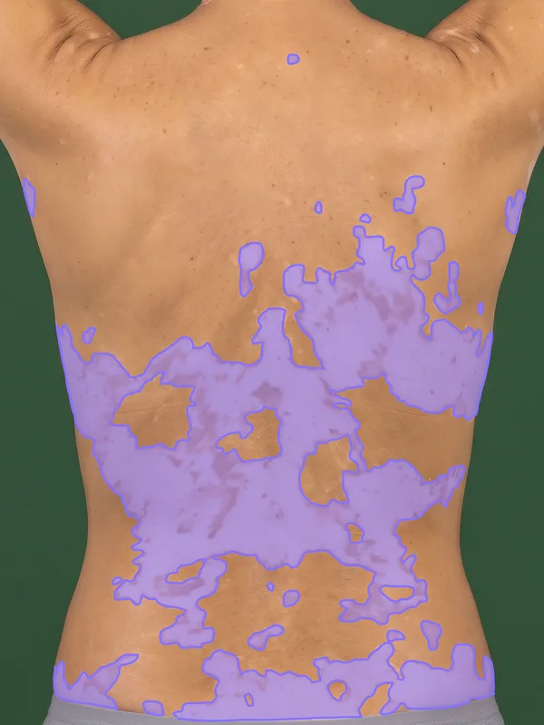
Back, 33% depigmented
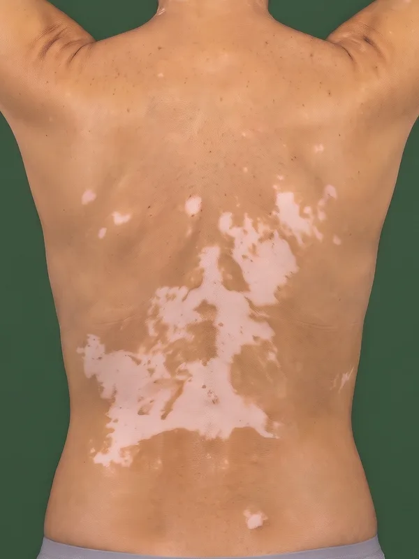
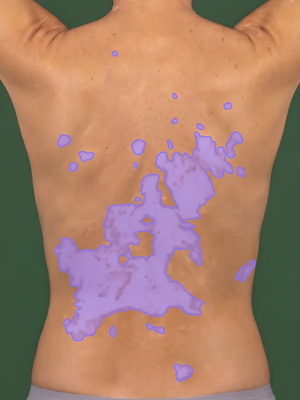
Back, 18% depigmented
Drag the slider to show or hide the depigmented skin the model found. Repigmentation shows as smaller lesions and as islands of pigment within them. Synthetic images, not patient records.
Manual vs. automated scoring
Manual scoring
VASI estimated by a clinician
- Extent per site: Estimated in palm units
- Degree of depigmentation: Graded per area, in seven steps
- Resolution: Coarse steps, often rounded
- Reproducibility: Depends on the rater and varies across sites
- Training: Calibration exercises to align raters
- Time per assessment: Full-body examination and estimation per visit
Automated scoring
Pixel-level segmentation of the standard photographs
- Extent per site: Measured from the pixels of each photograph
- Degree of depigmentation: Measured together with the extent; see how the automated score relates to the VASI
- Resolution: Continuous percentage
- Reproducibility: Same score for the same image, every time
- Training: Following the capture protocol instead of calibrating estimates
- Time per assessment: Seconds of AI analysis, plus capture of the standard views
The model version is locked at study initiation, so every patient is scored by the same model from first visit to last; see model version locking. The validation of the model is on Clinical evidence.
The AI reads depigmentation from what the photograph shows. Glare, very fair skin and other causes of light skin can look like vitiligo, and the model measures the extent of depigmentation without identifying its cause; see Limitations.