Scoring Methodology
This page provides the technical detail on how Legit.Health computes the automated PASI for psoriasis, from image input through to the final severity value.
The PASI (Psoriasis Area and Severity Index) is the severity score used in almost every psoriasis clinical trial. It combines two things that can both be read from a photograph: how much skin is affected, and how inflamed, thick and scaly that skin looks. This page explains each of those two measurements first, and then how the index combines them into a single number.
The example images on this page are synthetic dermatology imagery used for illustration; they are not patient records.
Affected body surface area
The first of the two measurements is extent: how much skin the disease covers. Estimating it by eye is the most operator-dependent step of manual scoring, and it is the input the AI measures most precisely.
How the AI measures affected area
A deep learning model analyses each full-body photograph in two passes.
First it locates the body and divides it into the four PASI regions, using anatomical landmarks so that every pixel is attributed to the right one. This step carries more weight than it appears to: the index counts each region differently, so skin credited to the wrong region changes the final score.
Then it classifies every pixel as affected skin, unaffected skin, or non-skin (background, clothing, hair). Counting those pixels gives the percentage of affected skin in each body region: the trunk might come out at 40% while the legs come out at 5%.
Combining the four regional percentages, weighted by how much of the body each region represents, gives the body surface area (BSA): one number for the whole patient, from 0 to 100%.
The PASI uses the four regional percentages, not the whole-body BSA, which is reported alongside it and used in its own right by treatment guidelines. What the index does with those percentages, and how it combines them with the intensity of the signs, is the subject of the two sections that follow.
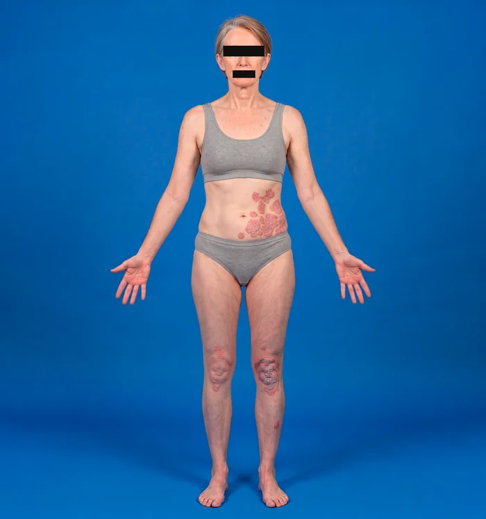
Input: body area photograph.
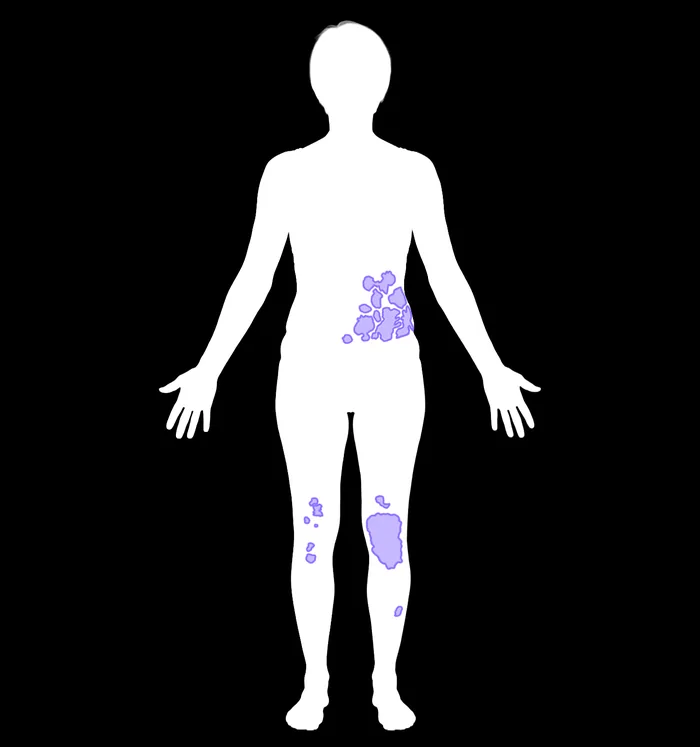
Output: affected areas detected across the body.
In manual scoring, a dermatologist estimates by eye what percentage of each region is covered by plaques. Dermatologists scoring the same patients differ by a mean of around 3.3 PASI points (Bożek et al., 2018), a gap that matters when a PASI of 10 is the threshold for systemic therapy.
Counting pixels removes that judgement. The same photograph always yields the same percentage, whoever submits it and whenever they do.
Intensity scoring: the three clinical signs
Alongside extent, the PASI grades the intensity of three disease signs in each body region, each on a 0 to 4 ordinal scale.
| Sign | Description |
|---|---|
| Erythema () | Redness of the plaque caused by inflammatory vasodilation, graded by how intense and widespread the red discoloration is. |
| Induration () | Thickness and elevation of the plaque above the surrounding skin, reflecting the degree of epidermal and dermal infiltration. |
| Desquamation () | Silvery-white scaling on the surface of the plaque, produced by the accelerated turnover of keratinocytes typical of psoriasis. |
In a live examination induration is usually assessed by palpating the plaque. The automated PASI grades it from the photograph alone, so no palpation is needed, and its grades agree with expert consensus at least as closely as dermatologists agree with each other. See production model performance.
Each sign is graded on the same five-point scale in every region:
| Grade | 0 | 1 | 2 | 3 | 4 |
|---|---|---|---|---|---|
| Level | None | Mild | Moderate | Severe | Very severe |
The three sign scores are summed per region, giving an intensity total of 0 to 12 for that region.
The PASI formula
The PASI asks the same two questions in each of four body regions: how much skin is affected, and how bad the affected skin looks. It multiplies those two answers together, weights the result by how much of the body that region represents, and adds the four regions up.
Each part of that product is explained below: the area score , the intensity total from the three signs above, and the region weight .
The area score: from a measured percentage to a 0 to 6 value
The PASI does not use the measured percentage directly. It sorts it into one of seven bands, and the band number is what enters the formula.
Area score by affected surface of the region
Each block is one area score, and its width is the share of the region it covers. Scores 1 and 6 span ten percentage points; scores 2 to 5 span twenty. A region with no affected skin scores 0.
The bands are not of equal width: scores 1 and 6 cover ten percentage points each, while scores 2 to 5 cover twenty. This coarseness belongs to the index, which was designed in 1978 for a clinician estimating by eye, not to the measurement.
A region at 31% affected and one at 49% both score 3. The AI measures the percentage exactly and the formula then rounds it away, so the gain is not a finer number: it is that the same photograph always lands in the same band, at every site and every visit. In a trial, that consistency matters more than the extra decimal place.
The region weight: how much each region counts
Each region carries a fixed weight, set by roughly how much of the body surface it represents. The weights never change and they add up to 1.
The four PASI regions and their fixed weights. These are anatomical proportions, not severity.
The affected area is how much of a given region is covered by plaques. It changes with the patient and with the visit, and it is what becomes the 0 to 6 score.
The region weight is fixed. The trunk always contributes 30% of the total, however much of it is affected.
A consequence worth knowing: the same plaque counts four times more on the legs than on the head.
A worked example
A patient has plaques on the trunk only, covering 40% of it. The other three regions are clear.
Trunk
That same patient has a whole-body BSA of 12%, because 40% of the trunk is 40% of 30% of the body. The two numbers answer different questions: the BSA counts only how much skin is involved, while the PASI also counts how inflamed, how thick and how scaly it is, and where on the body it sits.
The maximum PASI is 72, reached when all three signs score 4 in all four regions and every region is 90% affected or more.
Severity scale
Clear
Mild
Moderate
Severe
These are the severity bands of the rule of tens, the convention used in European treatment guidelines, where a PASI above 10 marks the threshold of moderate-to-severe disease and eligibility for systemic and biologic therapy.
Change over time, rather than the band at a single visit, is what a trial measures. The platform computes response against the patient's own baseline at every visit; see PASI response milestones.
Manual vs. automated scoring
Manual scoring
PASI graded by a clinician
- Affected area: Estimated by eye, then mapped to a 0 to 6 scale
- Sign scoring: Subjective, 0 to 4 per sign, 16 assessments per patient
- Reproducibility: Inter-rater variability across sites
- Time per assessment: Several minutes per patient
Automated scoring
Automated PASI computed by Legit.Health
- Affected area: Pixel-level segmentation, objective
- Sign scoring: A dedicated model per sign, validated against expert consensus
- Reproducibility: Identical score for identical image, every time
- Time per assessment: Under 2 seconds, plus 3 to 5 minutes of image capture
The model version is locked at study initiation, so every patient in a trial is scored by the same models from first visit to last; see model version locking. The measured agreement of each component model against dermatologist consensus is reported on Clinical evidence.
Because the platform scores from standardised photographs rather than a live examination, signs that a clinician might normally palpate, such as induration, are read from visual cues. Each model is accepted only when it is non-inferior to expert inter-rater variability on the same photographs, so the AI is at least as consistent as dermatologists working from the same images.