Scoring Methodology
The platform from Legit.Health scores hidradenitis suppurativa from lesion counts: the software detects every lesion, classifies it by type and scores the visit, and the investigator then reviews the result after examining the patient. The reported IHS4 and HiSCR are calculated from the confirmed counts, and the counts are reported in their own right, so any other count-based measure a protocol needs comes from the same capture.
Lesion detection and counting
- Photograph: Each affected region.
- Detect: A bounding box per lesion.
- Classify: Nodule, abscess or tunnel.
- Count: Three counts per visit.
- Score: Preliminary IHS4 and HiSCR.
- Review: The investigator confirms, edits or adds lesions.
- Recalculate: Final IHS4 and HiSCR.
A deep learning object detection model, trained on images annotated by a board of specialists and unified into a consensus reference, finds each lesion and assigns it to one of three types.
Inflammatory nodules
A firm, rounded, tender inflammatory lump, without pus. The earliest active lesion.
What the AI looks for: Solid, raised, well-defined borders, uniform colour
Abscesses
An inflamed lesion filled with pus, fluctuant to the touch. More active disease.
What the AI looks for: Fluctuant appearance, surrounding erythema, irregular surface
Draining tunnels
A tract under the skin that opens to the surface and can discharge pus or fluid.
What the AI looks for: Linear tracts, surface openings, discharge
Investigator review
Hidradenitis suppurativa is the one indication where the software's analysis is not the final word, because much of the disease sits beneath the skin and cannot be confirmed from a photograph alone. Once the software has returned its detections and scores, the investigator examines and palpates the patient and, in the annotation interface, confirms each detected lesion, corrects its type, removes it, or adds lesions the photograph does not show, before confirming the assessment.
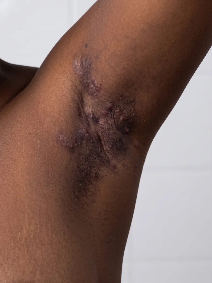
The affected region, captured at the visit
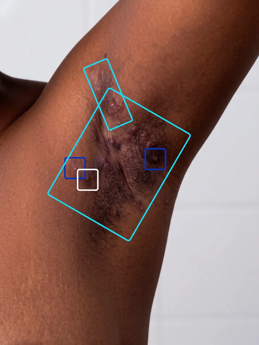
Each lesion detected and classified by the AI
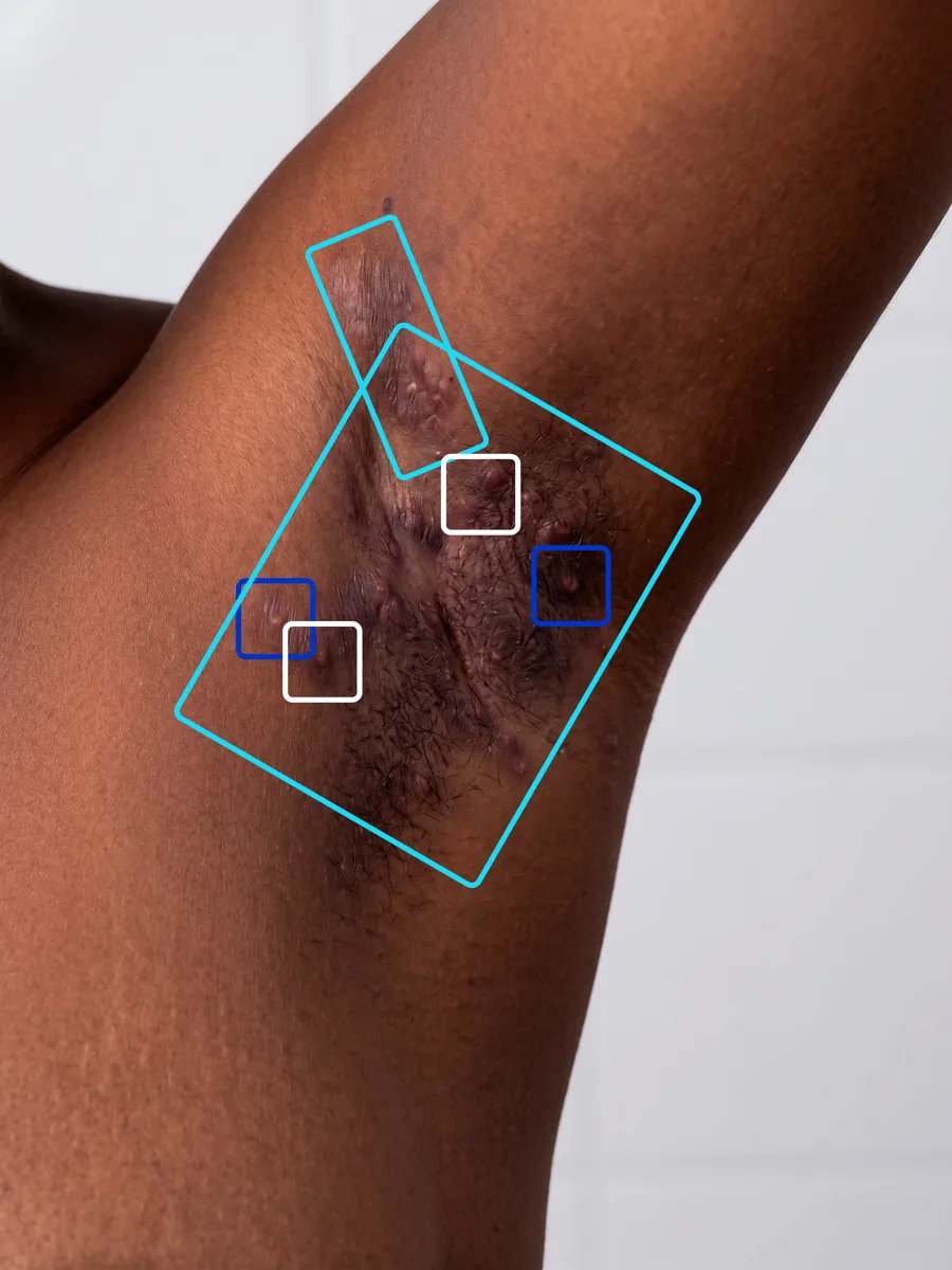
Detections confirmed, and a lesion added after palpation
The three counts for the visit (inflammatory nodules, abscesses and draining tunnels) are then recalculated from the confirmed lesions, together with the IHS4 and HiSCR. These confirmed values are the ones reported and exported.
Hidradenitis suppurativa develops mostly beneath the skin. A photograph shows a tunnel's opening and the tract it outlines, but whether it is actively draining depends on the patient's history and the clinical examination, which the image does not carry. The AI therefore counts every visible tunnel. See Limitations.
IHS4
The International Hidradenitis Suppurativa Severity Score System (IHS4) was developed by the European Hidradenitis Suppurativa Foundation and validated in 2017 (Zouboulis et al., British Journal of Dermatology). It is a dynamic score that follows the lesions present at each visit.
It weights each lesion type by how advanced the disease it reflects:
where each term is the number of lesions of that type counted at the visit.
IHS4 is purely a count: no affected area and no calibration markers, only every lesion visible and in focus. For the same reason the scale has no maximum, unlike PASI or EASI (72).
Severity thresholds
The bands are those defined in the original IHS4 validation study.
IHS4 severity bands. The severe band is open-ended because every additional lesion adds to the score.
Investigators disagree on which lesions are abscesses and which are nodules, and on whether a tract is a tunnel, and each disagreement moves the score by 1 to 4 points. The AI applies one classification boundary to every image, so every investigator starts from the same first read instead of counting from scratch.
HiSCR
The Hidradenitis Suppurativa Clinical Response (HiSCR) is the response endpoint most used in hidradenitis suppurativa trials. Where IHS4 scores one visit, HiSCR compares a follow-up visit with the patient's baseline visit, using the same lesion counts. It is built on the AN count:
A patient achieves HiSCR at a visit when all three conditions below are true. If any one fails, HiSCR is not achieved.
-
AN count falls by at least half
The AN count at the visit is 50% or less of the AN count at baseline
-
Abscesses do not increase
The number of abscesses is the same as at baseline or lower
-
Draining tunnels do not increase
The number of draining tunnels is the same as at baseline or lower
The platform stores the baseline counts and checks the three conditions automatically at every follow-up visit. Stricter versions used in some protocols, such as HiSCR 75 or HiSCR 90, raise the first condition to a 75% or 90% reduction.
A worked example
The same patient followed across a 48-week trial, with an assessment every 16 weeks. The IHS4 tracks how much disease remains at each visit; the AN count, measured against the baseline, shows when the patient reaches HiSCR 50 and HiSCR 75.
IHS4 by visit
AN count by visit
| Visit | Baseline | Week 16 | Week 32 | Week 48 |
|---|---|---|---|---|
| Inflammatory nodules | 8 | 4 | 2 | 1 |
| Abscesses | 4 | 1 | 1 | 0 |
| Draining tunnels | 2 | 2 | 2 | 1 |
| IHS4 | 24 Severe | 14 Severe | 12 Severe | 5 Moderate |
| AN count | 12 | 5 (−58%) | 3 (−75%) | 1 (−92%) |
| HiSCR 50 | Reference | Achieved | Achieved | Achieved |
| HiSCR 75 | Reference | Not yet | Achieved | Achieved |
The two measures move differently. The patient reaches HiSCR 50 at week 16 and HiSCR 75 at week 32, while the IHS4 stays in the severe band until the draining tunnels, which weigh most and respond slowest, start to close.
IHS4 and HiSCR
- Output
- A score from 0 upwards, with severity bands
- Needs baseline
- No
- Typical use
- Severity at inclusion, stratification, secondary endpoint
- Output
- Achieved or not achieved
- Needs baseline
- Yes
- Typical use
- Primary efficacy endpoint
IHS4 and HiSCR are calculated from the same confirmed lesion counts. The choice of primary and secondary endpoints is made during protocol design.
Manual vs. automated scoring
Manual scoring
IHS4 counted by a clinician
- Lesion identification: Visual inspection and palpation
- Type classification: Clinician judgement, abscess vs. nodule often ambiguous
- Reproducibility: Low inter-rater agreement across sites
- Training: Calibration exercises to align raters
- Time per assessment: Grows with lesion burden: every lesion found, examined and classified
Automated scoring
AI detection with investigator review
- Lesion identification: AI detection on every photograph, confirmed by the investigator
- Type classification: One consistent boundary, learned from specialist consensus
- Reproducibility: Same first read for the same image, every time
- Training: Reviewing detections instead of counting from scratch
- Time per assessment: Seconds of AI analysis, plus capture and the investigator's review
The model version is locked at study initiation, so every patient is scored by the same model from first visit to last; see model version locking. The peer-reviewed validation of the model is on Clinical evidence.
The AI reads the lesions from visual cues. What only touch can tell, such as the fluctuance of an abscess or involvement beneath the skin, is established by the investigator's examination during review; see Limitations.